Label the Skeleton
Tap a marker on the skeleton and pick the bone it belongs to. There are twelve markers, running from the skull down to the shin. The figure faces you, so the skeleton's own right side is on the left of the picture, and every arm and leg marker here sits on its left limbs.
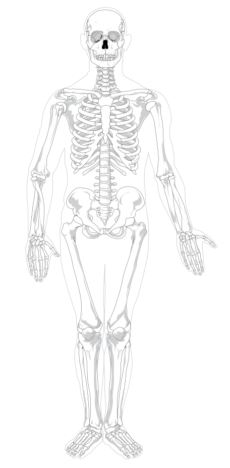
Pick the name of the highlighted marker.
All Nursing & Health quizzes
- Labeling quiz · 10 labels
Label the Heart Diagram
- Practice test · 15 questions
Anatomy and Physiology Practice Test
- Labeling quiz · 10 labels
Label the Neuron

Now make your own
Upload your prep book, slides or notes, or just tell Bo what you want to learn. You do not need a document to start.
All 12 labels on this diagram
Every marker on the figure is listed below with what it does. Answers stay collapsed until you open them, so you can quiz yourself as you scroll.
1. Skull
Show answer
The bony case around the brain, made of eight cranial bones fused at jagged seams called sutures. Everything above the eye sockets on this figure is cranium.
2. Mandible
Show answer
The lower jaw, and the only bone in the skull that moves. It carries the lower teeth and hinges at the temporomandibular joint just in front of each ear.
3. Clavicle
Show answer
The collarbone, running from the top of the sternum out to the shoulder. It props the shoulder away from the chest, and its shallow S shape is easy to spot on any front view.
4. Sternum
Show answer
The breastbone down the middle of the chest. The upper ribs join it through costal cartilage, which is why the rib cage can spring outwards when you breathe in.
5. Rib
Show answer
One of the twelve pairs that form the cage protecting the heart and lungs. The top seven pairs reach the sternum directly, three join the cartilage above them, and the bottom two are the floating ribs.
6. Humerus
Show answer
The single bone of the upper arm, from the shoulder joint to the elbow. It is the long bone people mean when they say they have broken an arm above the elbow.
7. Ulna
Show answer
The forearm bone on the little finger side. It is the thicker of the two at the elbow, where its hooked end forms the point you can feel when you bend your arm.
8. Radius
Show answer
The forearm bone on the thumb side. It is thin at the elbow and broad at the wrist, and it is the one that rotates over the ulna when you turn your palm downwards.
9. Pelvis
Show answer
The basin of bone carrying the weight of the upper body into the legs. Each side is three bones fused together, and the broad flared blade you can see here is the ilium.
10. Femur
Show answer
The thigh bone, the longest and strongest bone in the body. Its ball shaped head sits in the socket of the pelvis, and it angles inwards so the knees end up under the body's centre of gravity.
11. Patella
Show answer
The kneecap, a sesamoid bone sitting inside the tendon of the thigh muscles. It is not joined to the leg bones by a joint of its own, it rides over the front of the knee as it bends.
12. Tibia
Show answer
The shin bone, the thicker and inner of the two bones in the lower leg. It takes almost all the weight, which is why a broken tibia stops you walking and a broken fibula often does not.
Axial or appendicular, and why the split is worth knowing
An adult skeleton has around 206 bones, and no exam asks you to name them all. What exams do ask for is the split into two groups, because it organises everything else.
The axial skeleton is the core: the skull, the vertebral column, the ribs and the sternum. It protects the brain, the spinal cord, the heart and the lungs, and it barely moves. On this figure that is the head, the spine running down the middle, and the whole rib cage.
The appendicular skeleton is the limbs and the girdles that hang them on: the clavicle and scapula at the shoulder, the arm and hand, the pelvis, and the leg and foot. It is built for movement rather than protection. Of the twelve markers here, five are axial and seven are appendicular.
The number itself is worth a caveat. Babies are born with about 270 bones and many fuse during growth, and adults genuinely differ, so 206 is the usual figure rather than a fixed one.
The pairs that get mixed up, and how to tell them apart
Almost every mark lost on a skeleton diagram goes to one of three confusions.
Radius and ulna. The radius is on the thumb side, thin at the elbow and wide at the wrist. The ulna is on the little finger side, thick at the elbow and thin at the wrist. The memory hook most people keep is that the radius rotates, which is what lets you turn your palm over.
Tibia and fibula. The tibia is the thick inner bone that carries the weight, the fibula the thin outer one that mostly anchors muscle and forms the outer ankle bone. The fibula is visible on this figure as the slender bone beside the tibia, but it is deliberately not marked, because at this scale a marker on it would sit almost on top of the tibia's.
Left and right. The figure faces you, so its right side is on your left. Anatomical left and right always belong to the body being described, never to the person looking at it.
Two more structures worth finding while you are here, both unmarked: the scapula, the shoulder blade sitting mostly behind the rib cage and only partly visible from the front, and the vertebral column running down the centre, which you can see between the ribs and above the pelvis.
How StudyPDF turns your own diagrams into labeling practice
Twelve markers on one figure will get you through a quiz. They will not get you through a lab practical, where the diagram is the one from your own textbook, often a posterior view or a close up of a single region, with the labels your instructor used.
StudyPDF works from your material instead. Upload the chapter, the lecture slides or the lab manual, and Bo, the study agent, builds practice from those exact pages, including the figures in them. Every explanation cites where the answer came from, so you can go straight back to the page when something looks off.
You do not need a file to start. Name a topic instead, for example the bones of the axial skeleton or the bones of the hand, and Bo builds practice from that. Starting is free.
Written by the StudyPDF team. Last updated 2026-08-19.