Label the Eye Diagram
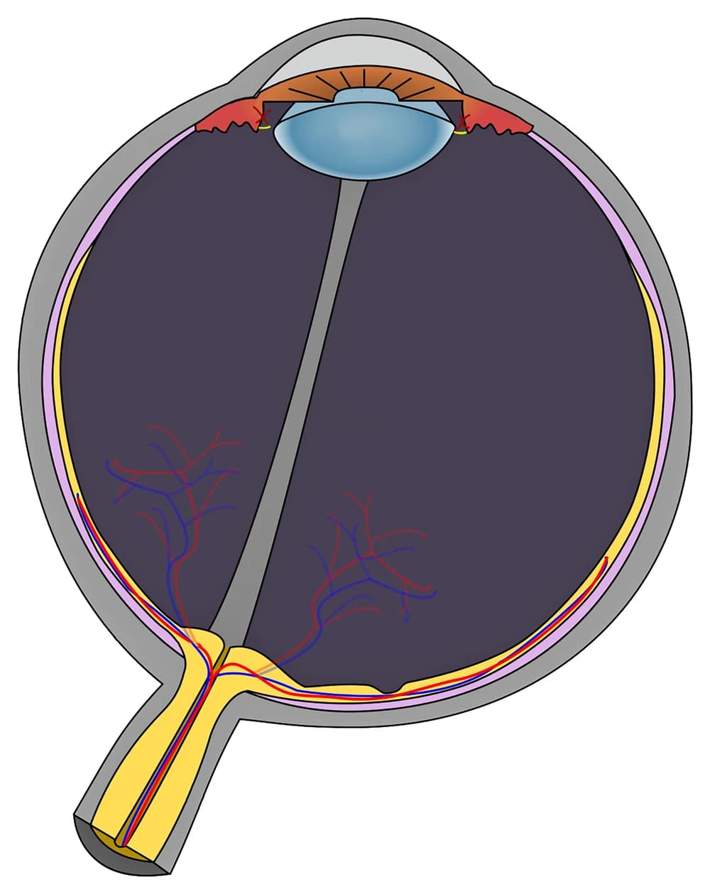
Tap a marker on the eye and pick the part it points to. There are twelve, from the cornea at the front to the optic nerve at the back. The figure shows the eye cut in half horizontally, so the front of the eye is at the top of the picture and light travels downwards through it to the retina.
Pick the name of the highlighted marker.
All Nursing & Health quizzes
- Labeling quiz · 10 labels
Label the Neuron
- Labeling quiz · 10 labels
Label the Heart Diagram
- Practice test · 15 questions
Anatomy and Physiology Practice Test

Now make your own
Upload your prep book, slides or notes, or just tell Bo what you want to learn. You do not need a document to start.
All 12 labels on this diagram
Every marker on the figure is listed below with what it does. Answers stay collapsed until you open them, so you can quiz yourself as you scroll.
1. Cornea
Show answer
The clear dome at the very front of the eye. It does most of the focusing, about 70 percent according to the American Academy of Ophthalmology, and the lens only fine tunes what the cornea has already bent. On this figure it is the curved band across the top, where the gray outer wall turns transparent.
2. Aqueous humor
Show answer
The watery fluid filling the front of the eye between the cornea and the lens, most of it in the anterior chamber between the cornea and the iris. The ciliary body makes it all the time and it drains away at the angle where the iris meets the cornea, and that balance is what keeps the pressure inside the eye steady.
3. Iris
Show answer
The colored ring you see when you look at someone's eye. It is a muscle: one set of fibers narrows the pupil in bright light and another widens it in the dark. In a cross-section it shows up as the two brown flaps lying in front of the lens.
4. Pupil
Show answer
The opening in the middle of the iris. It is not a structure at all, just a hole, which is why it looks black from the outside. Here it is the gap between the two halves of the iris, where the front of the lens shows through.
5. Lens
Show answer
The clear, flexible disc behind the pupil. Thin threads called zonules hold it to the ciliary body, and when the ciliary muscle contracts, those threads slacken and the lens rounds up to focus on something close. It stiffens with age, which is why reading glasses often arrive in your forties.
6. Ciliary body
Show answer
The red ring of tissue just behind the iris, on both sides of the lens. It does two jobs: its muscle changes the shape of the lens for near and far focus, and its lining produces the aqueous humor.
7. Sclera
Show answer
The tough outer wall of the eyeball, the part you see as the white of the eye. It keeps the eye's shape and gives the six muscles that move the eye something to pull on. At the front it continues into the cornea.
8. Choroid
Show answer
The thin layer of blood vessels between the sclera and the retina, shown here in pink. It feeds the outer layers of the retina, and its dark pigment soaks up stray light so it does not bounce around inside the eye.
9. Retina
Show answer
The light sensitive lining at the back of the eye, shown in yellow. Its rods handle dim light and its cones handle color and fine detail, and together they turn light into nerve signals. On the figure it is the innermost of the three layers in the wall.
10. Vitreous body
Show answer
The clear gel filling the big chamber between the lens and the retina, which is most of the eyeball. It holds the retina in place against the wall. The gray strip running through it on this figure is the hyaloid canal, a leftover from development.
11. Fovea
Show answer
The small pit in the retina almost straight behind the lens, at the center of the macula. It is packed with cones and has no rods at its center, so it gives the sharpest vision you have. Whatever you look at directly is focused here.
12. Optic nerve
Show answer
The cable of about a million nerve fibers carrying signals from the retina to the brain. The spot where it leaves the eye, the optic disc, has no rods or cones, which is what creates the blind spot in each eye.
Three layers, two fluids, one lens
The eye looks complicated on a diagram, but almost every part fits into a simple plan, and the plan is what exams test.
The wall of the eye has three layers. The outer layer is tough and protective: the sclera around most of the eyeball, turning into the clear cornea at the front. The middle layer carries the blood supply and the muscles: the choroid at the back, the ciliary body further forward, and the iris at the very front. The inner layer is the retina, the only part that actually detects light. On this figure you can see all three as stacked bands along the wall, gray, then pink, then yellow.
The inside is split by the lens. In front of it is the aqueous humor, a thin fluid that is made and drained all the time. Behind it is the vitreous body, a gel that stays put. The two are easy to tell apart once you remember that aqueous means watery and that it sits at the front, where it can flow.
Light follows the same order every time: cornea, aqueous humor, pupil, lens, vitreous body, retina. If you can recite that path, you can place most of the markers on any eye diagram without having seen it before.
The parts that get mixed up, and what is on the figure but not marked
Most lost marks on an eye diagram come from a few pairs.
Iris and pupil. The iris is the colored muscle, the pupil is the hole in it. A marker on the opening is always the pupil, a marker on the tissue around it is always the iris.
Cornea and sclera. They are one continuous outer wall, clear at the front and white everywhere else. The cornea is only the clear front window.
Choroid and retina. Both are thin layers inside the sclera. The choroid is the middle one and carries blood, the retina is the innermost and senses light. When in doubt, the retina is the layer touching the vitreous.
Fovea and optic disc. Both sit at the back. The fovea is the point of sharpest vision, the optic disc is where the nerve leaves and there is no vision at all. On this figure the optic disc is where the yellow optic nerve meets the wall, to the left of the fovea.
A few structures are visible but deliberately unmarked: the optic disc itself, the zonules (the short yellow threads holding the lens), the retinal blood vessels in red and blue, and the hyaloid canal, the gray strip running from the lens to the optic disc. The macula, the area around the fovea, is too small to show as its own region at this scale.
How StudyPDF turns your own diagrams into labeling practice
Twelve markers on one figure are a good start. A biology test or an anatomy practical will use the diagram from your own textbook or slides, often a front view of the eye or a close up of the retina, with the labels your teacher picked.
StudyPDF works from your material instead. Upload the chapter, the lecture slides or the worksheet. When your slides have a labeled diagram of the eye, you can turn it into image occlusion flashcards from the New material form: the labels are covered and you recall each one. Quizzes and practice tests come from the same pages, and every explanation cites where the answer came from. Bo, the tutor, explains any part you get wrong from your own pages.
You do not need a file to start. Name a topic instead, for example the parts of the eye and their functions, or how the eye focuses, and Bo sets it up so you can build practice from it. A free account covers about four new materials a week, and Pro and Ultra cover more.
Written by the StudyPDF team. Last updated 2026-08-19.