Label the Neuron
Tap a marker on the neuron and pick the part it belongs to. There are ten markers, running from the dendrites through the cell body and out along the axon. Two things about the drawing before you start: the pale grey cells in the background are neighbouring neurons and are not part of the one you are labeling, and the two circles are magnified views, one of a synapse and one of the myelin in cross section.
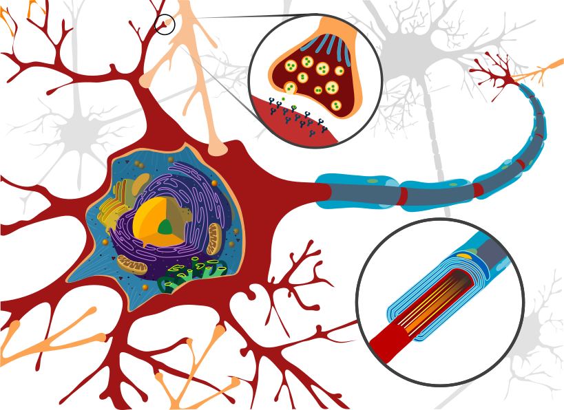
Pick the name of the highlighted marker.
All Nursing & Health quizzes
- Labeling quiz · 10 labels
Label the Heart Diagram
- Practice test · 15 questions
Anatomy and Physiology Practice Test
- Labeling quiz · 12 labels
Label the Skeleton

Now make your own
Upload your prep book, slides or notes, or just tell Bo what you want to learn. You do not need a document to start.
All 10 labels on this diagram
Every marker on the figure is listed below with what it does. Answers stay collapsed until you open them, so you can quiz yourself as you scroll.
1. Dendrite
Show answer
The branching extensions that receive signals from other neurons and carry them towards the cell body. A neuron usually has many of them, which is how one cell can listen to thousands of others at once.
2. Cell body
Show answer
Also called the soma or perikaryon. It holds the nucleus and the organelles, and it adds up the incoming signals from the dendrites to decide whether the neuron fires.
3. Nucleus
Show answer
Holds the neuron's DNA and directs protein synthesis. Neurons are unusually demanding cells, so the nucleus is large and the nucleolus inside it is prominent.
4. Nissl body
Show answer
Stacks of rough endoplasmic reticulum studded with ribosomes, named after the stain that first showed them. They make the neurotransmitters and membrane proteins the cell needs, and they are found in the cell body and dendrites but not in the axon.
5. Golgi apparatus
Show answer
Packages the proteins built by the Nissl bodies into vesicles. In a neuron much of that cargo travels the whole length of the axon to reach the terminals.
6. Mitochondrion
Show answer
Supplies the ATP the neuron runs on. Nerve cells are among the most energy hungry in the body, largely because pumping ions back across the membrane after every impulse costs so much.
7. Axon hillock
Show answer
The cone where the axon leaves the cell body. This is the trigger zone: if the summed signals reaching it cross threshold here, an action potential starts, and if they do not, nothing happens.
8. Myelin sheath
Show answer
The fatty insulation wrapped around the axon in segments. It is made by Schwann cells outside the brain and spinal cord and by oligodendrocytes inside them, and it speeds conduction up enormously.
9. Node of Ranvier
Show answer
The bare gap between two myelin segments. The impulse jumps from node to node instead of travelling along the whole membrane, which is called saltatory conduction and is why a myelinated axon is so much faster.
10. Axon
Show answer
The single long fibre carrying the impulse away from the cell body towards the terminals. A neuron has many dendrites but only one axon, and that axon can be over a metre long.
Follow the signal and the labels order themselves
A neuron is easier to label if you stop treating it as a shape and start treating it as a route. The signal only ever goes one way.
It arrives on the dendrites. It travels into the cell body, which adds up everything coming in. It reaches the axon hillock, the cone where the axon begins, and if the total crosses threshold an action potential fires. It then runs down the axon, jumping between the nodes of Ranvier where the myelin sheath is interrupted, until it reaches the terminals and crosses a synapse to the next cell.
That single sentence contains six of the ten markers in the right order. If you can recite the route, the only labels left are the organelles in the cell body: the nucleus, the Nissl bodies, the Golgi apparatus and the mitochondria.
What the two circles on the figure are showing you
The two circles are not structures. They are magnified views of parts of the same neuron, and they are worth reading because they answer the two questions this diagram usually raises.
The circle at the top magnifies a synapse. You can see the vesicles inside the terminal holding neurotransmitter, the gap itself, and the receptors on the other side. Nothing physically crosses from one neuron to the next: the electrical signal stops at the terminal, chemicals cross the gap, and a new signal starts on the far side.
The circle at the bottom right cuts through the axon to show what myelin actually is. It is not a coating painted on, it is a glial cell wrapped around the axon many times over, which is why it looks like layers rather than a single sleeve.
One more thing about the drawing: the pale grey cells behind the main neuron are other neurons, drawn to show that a real one is surrounded and contacted from every side. They are not labeled here, and neither are the peach coloured processes reaching in from off the figure, which belong to those neighbours.
How StudyPDF turns your own diagrams into labeling practice
One figure with ten markers will get you through a quiz. It will not get you through a lab practical or an exam where the diagram is the one from your own textbook, drawn from a different angle, with the labels your instructor used.
StudyPDF works from your material instead. Upload the chapter, the lecture slides or the lab manual, and Bo, the study agent, builds practice from those exact pages, including the figures in them. Every explanation cites where the answer came from, so you can go straight back to the page when something looks off.
You do not need a file to start. Name a topic instead, for example the action potential or saltatory conduction, and Bo builds practice from that. Starting is free.
Written by the StudyPDF team. Last updated 2026-08-19.