Label the Animal Cell
Tap a marker on the animal cell and pick the organelle it belongs to. There are ten markers, covering the boundary of the cell, the nucleus and the organelles a biology course asks you to name. Work outside in if you get stuck: the membrane and the cytoplasm are the easy two, and everything else sits inside them.
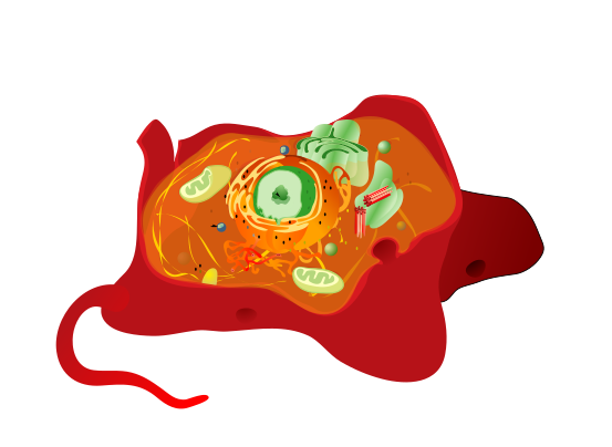
Pick the name of the highlighted marker.
All High School & Admission quizzes
- Labeling quiz · 10 labels
Label the Plant Cell
- Practice test · 15 questions
ASVAB Practice Test
- Practice test · 15 questions
GED Practice Test

Now make your own
Upload your prep book, slides or notes, or just tell Bo what you want to learn. You do not need a document to start.
All 10 labels on this diagram
Every marker on the figure is listed below with what it does. Answers stay collapsed until you open them, so you can quiz yourself as you scroll.
1. Cell membrane
Show answer
The outer boundary of the cell, also called the plasma membrane. It is a double layer of phospholipids that decides what gets in and out. An animal cell has no cell wall outside it, which is the difference examiners ask about most.
2. Cytoplasm
Show answer
The jelly filling the cell between the membrane and the nucleus. The organelles sit in it and most of the cell's chemical reactions happen in it. The watery part on its own is called the cytosol.
3. Nucleus
Show answer
The control centre. It holds the cell's DNA and decides which proteins get made, which is why it is usually the largest structure you can see. Red blood cells are the famous exception: mature ones have lost theirs.
4. Nucleolus
Show answer
The dense spot inside the nucleus. It is where ribosomes are built before they are shipped out into the cytoplasm. It is a region rather than a membrane bound organelle, so it has no envelope of its own.
5. Nuclear envelope
Show answer
The double membrane wrapped around the nucleus. It is pierced by nuclear pores that let messenger RNA out and proteins in, so the nucleus stays separate from the cytoplasm without being sealed off.
6. Rough endoplasmic reticulum
Show answer
The folded sheets running off the nuclear envelope. It looks rough because it is studded with ribosomes, the black dots on the figure, and those ribosomes make the proteins that get exported from the cell.
7. Smooth endoplasmic reticulum
Show answer
The tube network with no ribosomes on it. It makes lipids, stores calcium and breaks down toxins, which is why liver cells carry so much of it. Smooth and rough are the same network, just different jobs.
8. Golgi apparatus
Show answer
The stack of flattened sacs. It takes proteins from the endoplasmic reticulum, finishes them off and packages them into vesicles for delivery. The usual shorthand is the post office of the cell.
9. Mitochondrion
Show answer
The bean shaped organelle with the folded inner membrane. Aerobic respiration happens on those folds, so this is where most of the cell's ATP comes from. Muscle cells are packed with them.
10. Centrioles
Show answer
The pair of short cylinders near the nucleus. They organise the spindle fibres that pull chromosomes apart during cell division. Plant cells do not have them, which is another difference exams like.
The order to learn an animal cell in
Ten organelles at once is a lot to hold. It gets much easier if you learn them as three groups instead of one list.
The first group is the boundary and the filling: the cell membrane on the outside and the cytoplasm inside it. The second is the nucleus and everything attached to it: the nucleus itself, the nucleolus inside it, the nuclear envelope around it, and the rough endoplasmic reticulum that runs straight off that envelope. The third is the working organelles floating in the cytoplasm: smooth endoplasmic reticulum, Golgi apparatus, mitochondria and centrioles.
Grouped that way the diagram tells a story rather than being ten separate facts. The nucleus writes the instructions, the rough endoplasmic reticulum and its ribosomes build the protein, the Golgi apparatus packages it, and the mitochondria pay for all of it in ATP.
What animal cells have that plant cells do not
Most exam questions on this diagram are really comparison questions, so it is worth knowing the differences cold.
An animal cell has centrioles and a plant cell normally does not. An animal cell has no cell wall, no chloroplasts and no large central vacuole. Its shape is irregular, held by the cytoskeleton rather than by a rigid wall, which is why the figure above has that lumpy outline instead of the neat box a plant cell is drawn as.
What both have in common is the bigger list: cell membrane, cytoplasm, nucleus, nucleolus, ribosomes, both kinds of endoplasmic reticulum, Golgi apparatus and mitochondria. If a question asks what a plant cell has that an animal cell lacks, the answer is the wall, the chloroplasts and the central vacuole, in that order of how often it is asked.
One more thing worth noticing on the figure: the ribosomes are the black dots on the rough endoplasmic reticulum. They are not marked here because they are far too small to tap, but they are the reason that part of the network is called rough, and free ribosomes float in the cytoplasm too.
How StudyPDF turns your own diagrams into labeling practice
One figure with ten markers will get you through a quiz. It will not get you through a test where the diagram is the one in your own textbook, drawn from a different angle, with the labels your teacher actually used.
StudyPDF works from your material instead. Upload the chapter, the lecture slides or the revision guide, and Bo, the study agent, builds practice from those exact pages, including the figures in them. Every explanation cites where the answer came from, so you can go straight back to the page when something looks off.
You do not need a file to start. Name a topic instead, for example organelles and their functions or the difference between plant and animal cells, and Bo builds practice from that. Starting is free.
Written by the StudyPDF team. Last updated 2026-08-19.