Pick the name of the highlighted marker.
Keep practicing

Now make your own
Upload your prep book, slides or notes, or just tell Bo what you want to learn. You do not need a document to start.
About this diagram
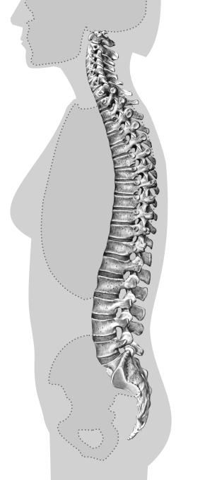
Tap a marker on the spine and pick the name it belongs to. There are ten, from the atlas at the top of the neck to the coccyx at the bottom. The figure is a side view, so the front of the body is on the left and the spinous processes point to the right.
All 10 labels on this diagram
Every marker on the figure is listed below with what it does. Answers stay collapsed until you open them, so you can quiz yourself as you scroll.
1.Atlas (C1)
The first cervical vertebra, the one that holds up the skull, which is how it got the name of the giant who carried the sky. It is a ring with no vertebral body and no real spinous process, and the rounded knobs at the base of the skull rest in its upper surfaces. That joint is where you nod yes.
2.Axis (C2)
The second cervical vertebra. A peg of bone called the dens rises from its body into the ring of the atlas, and the atlas and skull turn around that peg when you shake your head no. Its spinous process is thick and forked at the tip.
3.Cervical vertebrae
The seven vertebrae of the neck, C1 to C7. They are the smallest of the movable vertebrae, with short spinous processes, and the only ones with a small hole in each side process, the transverse foramen, that carries a vertebral artery on its way up to the brain. The marker sits on the body of one of the middle ones.
4.Vertebra prominens (C7)
The seventh cervical vertebra has the longest spinous process of the neck, and it is the bump you can feel at the base of the neck when you tip your head forward. The first thoracic vertebra can stick out almost as far, so the bump is a landmark and not proof. The marker sits on the process itself.
5.Thoracic vertebrae
The twelve vertebrae of the chest, T1 to T12. Each has small facets on its sides where a rib joins, which is why they are the only vertebrae that carry ribs. Their bodies get larger going down, and their long spinous processes slope steeply downward, like roof tiles. The marker sits on the body of one in the middle of the chest.
6.Intervertebral disc
The pad between the bodies of two neighboring vertebrae: a tough outer ring, the annulus fibrosus, around a soft gel center, the nucleus pulposus. It works as a shock absorber and lets the spine bend. The column is usually counted as having 23 of them, none between the atlas and axis and none inside the sacrum or coccyx.
7.Lumbar vertebrae
The five vertebrae of the lower back, L1 to L5. They have the largest and thickest bodies in the column because they carry the most weight, and short, blunt, boxy spinous processes that point straight back. They carry no ribs. The marker sits on the body of one of the middle ones.
8.Intervertebral foramen
The opening between two neighboring vertebrae through which a spinal nerve leaves the spinal canal. The pedicles of the vertebrae form its roof and floor, the bodies and the disc form its front, and the joints between the facets form its back. A bulging disc or a bone spur can narrow it and press on the nerve. It shows here as the pale oval gap.
9.Sacrum
Five vertebrae fused into one triangular bone, S1 to S5. It wedges between the two hip bones at the sacroiliac joints and forms the back wall of the pelvis. The fusion finishes in early adulthood, which is why the sacrum is counted as one bone in adults.
10.Coccyx
The tailbone at the bottom of the column, usually four small vertebrae fused together, though three to five is normal. It carries almost no weight when you stand, it is an anchor for some pelvic floor muscles and ligaments, and it can share some of the load when you sit leaning back.
33 vertebrae, five regions and the numbers to know
The vertebral column is counted as 33 vertebrae in five regions: 7 cervical in the neck, 12 thoracic in the chest, 5 lumbar in the lower back, 5 sacral and usually 4 coccygeal. The classic memory aid is 7, 12, 5, as in breakfast at 7, lunch at 12, dinner at 5. The sacral vertebrae fuse into one bone and the coccygeal ones into another, which leaves 24 separate, movable vertebrae plus the sacrum and coccyx, and is why an adult spine is also described as 26 bones.
This figure marks ten things: the three most-tested individual vertebrae (atlas, axis and C7), the five regions, one disc and one foramen. The original drawing came with colored bands that tint each region. We took them off, so you have to recognize each region by its shape. Cervical vertebrae are small and delicate, thoracic ones have long spinous processes that slope down, lumbar ones are the big boxy ones at the bottom, and the sacrum is the single triangular bone.
Seen from the side, the column is not straight. It has four curves: the neck and the lower back curve inward, which is called lordosis, and the chest and the sacrum curve outward, which is called kyphosis. The chest and sacral curves are there from birth. The neck curve develops as a baby starts to hold up the head, and the lower back curve as a child starts to sit up and stand.
Landmarks and the mix-ups that cost marks
The pairs below catch most people. Atlas and axis: the atlas is the top ring that holds the skull and has no body, while the axis is the second one, with the peg called the dens. Disc and foramen: the disc is the soft pad in front, between two bodies, and the foramen is the gap behind it that the spinal nerve passes through. Sacrum and coccyx: the sacrum is the large fused triangle and the coccyx is the small tail at its tip.
Two habits help. First, count from a landmark you can trust. The atlas is always the top one, so count down from it and treat the C7 bump as a hint, because on some people it is really T1. Second, read the processes. A long, sloping spinous process says thoracic, a short blunt one says lumbar, and a forked tip points to the middle of the neck.
Why are the curves not marked? A curve is a shape of the whole column, not a part you can point at, and any marker would sit on the same bones as a region marker. They are covered in the section above, and anatomy exams usually ask about them in words, not on a diagram.
How StudyPDF turns your own diagrams into labeling practice
Ten markers on one figure are a good start. A lab practical is harder: the spine is often a real one or the figure from your own textbook, it may be a single vertebra seen from above, and the instructor may ask for processes, facets and foramina as well as whole bones.
StudyPDF works from your material instead. Upload the chapter, the lecture slides or the lab manual. When your slides have a labeled diagram of the spine, you can turn it into image occlusion flashcards from the New material form: the labels are covered and you recall each one. Quizzes and practice tests come from the same pages, and every explanation cites where the answer came from. Bo, the tutor, explains any part you get wrong from your own pages.
You do not need a file to start. Name a topic instead, for example the parts of a typical vertebra or the spinal nerves, and Bo sets it up so you can build practice from it. Starting is free.
Written by the StudyPDF team. Last updated August 19, 2026.
Good to know
Is this vertebrae labeling quiz free?
Yes. All ten markers, your score and the explanation for every one are free to play in your browser. The printable label list needs a free StudyPDF account.
How many vertebrae are there in the spine?
Usually 33: 7 cervical, 12 thoracic, 5 lumbar, 5 sacral and about 4 coccygeal. In an adult the sacral ones are fused into one bone and the coccygeal ones into another, so 24 vertebrae are separate and movable. Three to five coccygeal vertebrae is a normal range.
How do I tell the atlas from the axis?
The atlas is C1, the top ring that carries the skull, and it has no vertebral body. The axis is C2, directly below, and it has a peg of bone, the dens, that points up into the atlas. The atlas lets you nod yes and the axis lets you shake your head no.
Why are the cervical and lumbar curves not marked on the diagram?
Because a curve is the shape of a whole region, not a single part you can point at, and a marker for it would sit on the same bones as the region marker. The curves are explained on this page instead: the neck and lower back curve inward, the chest and sacrum curve outward.