Label the Skull
Tap a marker on the skull and pick the bone it belongs to. There are eleven, one for every bone shown in its own color, from the frontal bone of the forehead to the mandible. The skull faces left, so you are looking at its left side, and markers 6 and 7 sit close together inside the eye socket.
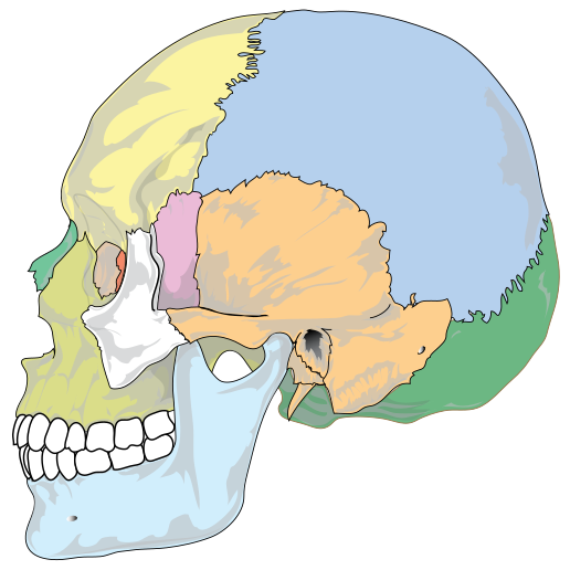
Pick the name of the highlighted marker.
All Nursing & Health quizzes
- Labeling quiz · 12 labels
Label the Skeleton
- Labeling quiz · 12 labels
Label the Eye Diagram
- Practice test · 15 questions
Anatomy and Physiology Practice Test

Now make your own
Upload your prep book, slides or notes, or just tell Bo what you want to learn. You do not need a document to start.
All 11 labels on this diagram
Every marker on the figure is listed below with what it does. Answers stay collapsed until you open them, so you can quiz yourself as you scroll.
1. Frontal bone
Show answer
The bone of the forehead, which also forms the roof of each eye socket. It meets the two parietal bones at the coronal suture, the wavy seam running over the top of the head behind it.
2. Parietal bone
Show answer
One of a pair that makes up most of the roof and the upper sides of the skull. The two parietal bones meet each other along the sagittal suture on top of the head, which this side view cannot show.
3. Temporal bone
Show answer
The bone at the side of the head, below the parietal. It houses the middle and inner ear, and the dark opening just behind the jaw joint on this figure is the ear canal, the external acoustic meatus. The mandible hinges against it at the temporomandibular joint.
4. Occipital bone
Show answer
The back and base of the skull. The spinal cord passes through its large opening, the foramen magnum, and the two occipital condyles beside that opening rest on the first vertebra, the atlas.
5. Sphenoid bone
Show answer
A single bat-shaped bone that runs right across the skull base. From the side you only see its greater wing at the temple, but it touches every other cranial bone, and a hollow on its upper surface, the sella turcica, holds the pituitary gland.
6. Ethmoid bone
Show answer
A light, spongy bone between the eye sockets that forms the roof and much of the upper walls of the nasal cavity. From the side you only see a thin plate of it in the inner wall of the eye socket, the narrow red strip under this marker. The nerves for smell pass through its cribriform plate.
7. Lacrimal bone
Show answer
The smallest bone of the face, a thin plate in the inner wall of the eye socket, in front of the ethmoid. A groove in it holds the lacrimal sac, which drains tears down into the nose.
8. Nasal bone
Show answer
One of two small bones side by side that form the bony bridge of the nose. Only the upper part of the nose is bone. The rest is cartilage, which is why a skull has an open hole where the tip of the nose would be.
9. Zygomatic bone
Show answer
The cheekbone, forming the outer wall and part of the floor of the eye socket. Its back end joins a projection of the temporal bone, and the two together make the zygomatic arch you can feel in front of your ear.
10. Maxilla
Show answer
The upper jaw, which carries the upper teeth. The two maxillae meet in the middle, form most of the hard palate and the floor of each eye socket, and each one holds the largest of the air-filled sinuses, the maxillary sinus.
11. Mandible
Show answer
The lower jaw, the largest and strongest bone of the face and the only skull bone that moves. Its upright back part, the ramus, rises to the joint with the temporal bone just in front of the ear canal.
Twenty-two bones, and the eleven you can see from the side
An adult skull is usually counted as 22 bones, split into two groups. The 8 cranial bones form the case around the brain: one frontal, two parietal, two temporal, one occipital, one sphenoid and one ethmoid. The 14 facial bones build the face: two nasal, two maxillae, two zygomatic, two lacrimal, two palatine, two inferior nasal conchae, one vomer and one mandible.
A side view shows eleven of those names, and they are the eleven markers here. Six are cranial: frontal, parietal, temporal, occipital, sphenoid and ethmoid. Five are facial: lacrimal, nasal, zygomatic, maxilla and mandible. Paired bones only appear once, because the other one is on the far side of the skull.
The palatine bones, the vomer and the inferior nasal conchae are missing for a simple reason: they sit deep inside the nose and mouth and cannot be seen from the side. The ear ossicles and the hyoid bone in the neck are not counted in the 22 either, which is why some textbooks give larger totals.
Sutures, landmarks and the mix-ups that cost marks
The cranial bones are locked together by sutures, jagged seams that are flexible in a baby and fuse slowly with age. Three are visible from the side. The coronal suture runs between the frontal and parietal bones, the lambdoid suture between the parietal and occipital bones, and the squamous suture curves along the top of the temporal bone. The fourth big one, the sagittal suture between the two parietal bones, runs along the top of the head and only shows from above.
One landmark comes up on almost every exam: the pterion, the small area at the temple where the frontal, parietal, temporal and sphenoid bones meet. It is one of the thinnest parts of the skull wall, with the middle meningeal artery running just underneath it. On this figure it is around the top of the pink sphenoid.
The errors people make on this diagram are predictable. Sphenoid and temporal get swapped because both show at the side of the head; the sphenoid is the narrow strip in front, the temporal the broad plate behind. The zygomatic arch is two bones, not one, the zygomatic in front and a projection of the temporal behind. In the eye socket, the lacrimal is the plate in front and the ethmoid sits behind it. And the maxilla is the upper jaw, fixed to the skull, while the mandible is the lower jaw that moves.
How StudyPDF turns your own diagrams into labeling practice
Eleven markers on one figure will get you through a quiz. A lab practical is harder: the skull is often a real one or the figure from your own textbook, it may be a front view or the skull base, and the instructor may ask for foramina and processes as well as whole bones.
StudyPDF works from your material instead. Upload the chapter, the lecture slides or the lab manual, and Bo, the study agent, builds practice from those exact pages, including the figures in them. Every explanation cites where the answer came from, so you can go straight back to the page when something looks off.
You do not need a file to start. Name a topic instead, for example the cranial bones and sutures or the foramina of the skull base, and Bo builds practice from that. Starting is free.
Written by the StudyPDF team. Last updated 2026-08-19.