Keep practicing

Now make your own
Upload your prep book, slides or notes, or just tell Bo what you want to learn. You do not need a document to start.
About this diagram
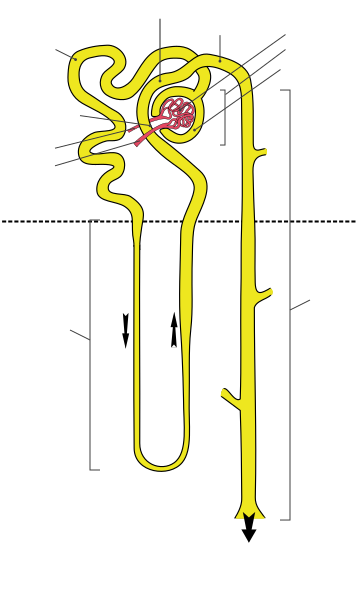
Tap a marker on the nephron and pick the part it belongs to. There are eleven markers: the filter, the two arterioles, the tubule from start to finish, and the cortex and medulla it runs through. The gray lines, brackets and arrows in the drawing are part of the artwork and do not name anything, and the dashed line marks where the cortex ends and the medulla begins.
All 11 labels on this diagram
Every marker on the figure is listed below with what it does. Answers stay collapsed until you open them, so you can quiz yourself as you scroll.
1.Glomerulus
The red tuft of capillaries where blood is filtered. Pressure pushes water, salts, glucose and other small molecules out of the blood into the capsule around it, while blood cells and large proteins stay behind. The fluid that gets through is called filtrate.
2.Bowman's capsule
The cup shaped wall around the glomerulus, drawn here as the yellow ring around the red tuft, with the marker on the bottom of the ring. It catches the filtrate and passes it into the tubule. The glomerulus and its capsule together are called the renal corpuscle.
3.Afferent arteriole
The vessel that brings blood to the glomerulus, and the thicker of the two red vessels in the drawing. Afferent arrives, efferent exits. Blood comes in through a wider vessel than the one it leaves by, which keeps the pressure in the tuft high.
4.Efferent arteriole
The thinner red vessel, which carries blood away from the glomerulus. Because it is narrower than the afferent arteriole, it holds the pressure inside the tuft up. It does not lead straight to a vein: it breaks up into a second set of capillaries that wrap around the tubules.
5.Proximal convoluted tubule
The first, tightly coiled stretch of tubule after the capsule, found in the cortex. It takes back most of what the body wants to keep: about two thirds of the filtered water and sodium, and nearly all the glucose and amino acids.
6.Descending limb (loop of Henle)
The thin tube that dives down into the medulla, the first leg of the loop of Henle. Water leaves it as it passes through ever saltier surroundings, so the fluid inside becomes more concentrated toward the turn at the bottom. The black arrow beside it shows the direction of flow.
7.Ascending limb (loop of Henle)
The tube that climbs back up toward the cortex after the turn. Unlike the descending limb it lets almost no water through, and salt is moved out of it, so the fluid gets more dilute as it rises. That salt is also what keeps the medulla salty enough for the descending limb to work.
8.Distal convoluted tubule
The second coiled stretch, back in the cortex, which here curls round past the glomerulus. Where the tubule comes back to touch the arterioles, a patch of cells called the macula densa senses the salt in the fluid and helps tune filtration. Hormones such as aldosterone and parathyroid hormone fine tune what it reabsorbs.
9.Collecting duct
The long straight tube that many nephrons drain into, running down through the medulla toward the renal pelvis. It makes the last adjustment to the urine: when antidiuretic hormone is high it lets water back into the body and the urine comes out concentrated.
10.Renal cortex
The outer layer of the kidney, above the dashed line. It holds the glomeruli and capsules and both convoluted tubules, so this is where filtration happens. The marker sits in the empty space beside the figure because it names a region, not a tube.
11.Renal medulla
The inner part of the kidney, below the dashed line. In this drawing it holds the loop of Henle and the collecting duct, and it is where the urine gets concentrated. In a whole kidney the medulla is arranged in cone shaped renal pyramids.
Follow the filtrate and the labels order themselves
A nephron is easier to label as a route than as a shape, because the fluid only moves one way and almost every marker sits on that route.
Blood arrives through the afferent arteriole and is filtered in the glomerulus. The fluid that is pushed out, called filtrate, is caught by Bowman's capsule and passes into the tubule: first the proximal convoluted tubule, then down the descending limb of the loop of Henle, round the turn and up the ascending limb, through the distal convoluted tubule and into the collecting duct, which carries it on toward the renal pelvis. The blood that was not filtered leaves the glomerulus through the efferent arteriole.
That is nine of the eleven markers in order. The other two, the renal cortex and the renal medulla, are places rather than tubes. In this drawing the cortex holds the filter and both coiled tubules, while the loop of Henle dips into the medulla and the collecting duct runs down through it. To remember the arterioles, afferent arrives and efferent exits.
For scale, each kidney holds roughly a million nephrons. Together they filter on the order of 180 liters of fluid a day, and only about 1 to 2 liters of that leaves the body as urine. Everything else is taken back.
Reading this figure: what the lines, colors and brackets mean
The drawing has more in it than eleven structures, and the extras are easy to mistake for part of the answer. Red is blood: the tangled tuft is the glomerulus and the two red tubes joining it are the arterioles. Yellow is the tubule. The black arrows show which way the fluid flows, down the first leg of the loop and up the second, and the big arrow at the bottom points on to the renal pelvis. The short stubs on the collecting duct are where other nephrons would drain in.
The gray lines and brackets are part of the original artwork and are not markers. The short bracket beside the capsule marks the renal corpuscle, meaning the glomerulus plus its capsule. The bracket on the left marks the loop of Henle and the long bracket on the right marks the collecting duct. The dashed line across the middle separates the cortex above from the medulla below, which is why those two markers sit in the empty space at the left, one above the line and one below it.
Two more things. The short tube at the top right that leads into the collecting duct is the connecting tubule, and it has no marker here. And this is a diagram, not a drawing to scale: a real nephron is microscopic, and its tubes are far longer and more tightly coiled than they look here.
How StudyPDF turns your own diagrams into labeling practice
One figure with eleven markers will get you through a quiz. A physiology exam or a lab practical will use the nephron diagram from your own course, drawn in a different style, with the terms your instructor expects.
StudyPDF works from your material instead. Upload the chapter, the lecture slides or your lab manual, and Bo, the study agent, builds practice from those exact pages, including the figures in them. Every explanation cites where the answer came from, so you can go straight back to the page.
You do not need a file to start. Name a topic instead, for example urine formation or the countercurrent mechanism, and Bo sets up practice on it. Starting is free.
Written by the StudyPDF team. Last updated August 19, 2026.
Good to know
What are the parts of the nephron, in order?
Following the fluid: the renal corpuscle (the glomerulus inside Bowman's capsule), the proximal convoluted tubule, the loop of Henle with its descending and ascending limbs, the distal convoluted tubule, and then the collecting duct. Strictly, the collecting duct is not part of the nephron because many nephrons drain into it, but it is nearly always taught and labeled with it.
What is the difference between the afferent and efferent arteriole?
Direction and width. The afferent arteriole brings blood into the glomerulus and the efferent arteriole carries it out. The efferent one is narrower, and that squeeze keeps the pressure inside the glomerulus high enough to push fluid out into the capsule.
Where do filtration, reabsorption and secretion happen?
Filtration happens only at the glomerulus. Reabsorption starts in the proximal tubule, which takes back most of the water, sodium, glucose and amino acids, and carries on in the loop of Henle, the distal tubule and the collecting duct. Secretion, where the tubule adds substances such as hydrogen ions, potassium and some drugs to the filtrate, happens along the proximal and distal tubules and the collecting duct.
Is this free, and is there an answer key I can print?
Yes. All eleven markers, your score and the explanation for every part are free to play in your browser. The printable label list needs a free StudyPDF account.