Label the Respiratory System
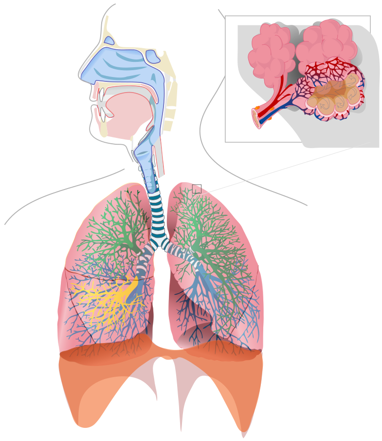
Tap a marker on the diagram and pick the part it belongs to. There are eleven markers, following the air from the nasal cavity down to the alveoli and the diaphragm. The head is drawn from the side and the chest from the front, so the right lung is on the left of the picture. The square at the top right is a magnified view of the tiny square drawn on the left lung.
Pick the name of the highlighted marker.
All Nursing & Health quizzes
- Labeling quiz · 10 labels
Label the Heart Diagram
- Practice test · 15 questions
Anatomy and Physiology Practice Test
- Practice test · 15 questions
TEAS Science Practice Test

Now make your own
Upload your prep book, slides or notes, or just tell Bo what you want to learn. You do not need a document to start.
All 11 labels on this diagram
Every marker on the figure is listed below with what it does. Answers stay collapsed until you open them, so you can quiz yourself as you scroll.
1. Nasal cavity
Show answer
The air space behind the nose. It warms, moistens and filters the air before it goes any deeper. The curved ridges drawn inside it are the nasal conchae, which stir the air so more of it touches the moist lining.
2. Pharynx
Show answer
The throat, a muscular tube behind the nose and mouth. Air and food share it, and they split at its lower end: air goes forward into the larynx and food goes back into the esophagus.
3. Epiglottis
Show answer
The flap of cartilage above the entrance to the larynx. When you swallow it folds down over the opening, so food and drink go into the esophagus instead of the airway.
4. Larynx
Show answer
The voice box, which sits between the pharynx and the trachea. It holds the vocal folds, which vibrate to make sound, and its largest cartilage, the thyroid cartilage, is the Adam's apple you can feel in the neck.
5. Trachea
Show answer
The windpipe, which carries air from the larynx down into the chest. C-shaped rings of cartilage, the stripes on the drawing, hold it open so it never collapses when you breathe in.
6. Main bronchus
Show answer
One of the two tubes the trachea splits into, one for each lung. The marker is on the left main bronchus. The right one is shorter, wider and more vertical, which is why inhaled objects end up on the right more often.
7. Right lung
Show answer
The larger of the two lungs, on the left of this picture because the figure faces you. It has three lobes, superior, middle and inferior, separated by two fissures you can see as thin lines.
8. Middle lobe
Show answer
The lobe only the right lung has, between the horizontal and oblique fissures. On this drawing its airways are colored yellow, which makes it easy to pick out.
9. Left lung
Show answer
The smaller lung, with two lobes instead of three. It gives up room for the heart, which leaves a curved dent in its front edge called the cardiac notch.
10. Alveoli
Show answer
The tiny air sacs at the end of the smallest airways, shown magnified in the inset. Their walls are one cell thick and wrapped in capillaries, so oxygen passes into the blood and carbon dioxide passes out right here.
11. Diaphragm
Show answer
The dome-shaped muscle under the lungs and the main muscle of breathing. When it contracts it flattens, the chest gets bigger and air flows in. When it relaxes it domes up again and air flows out.
Follow one breath and the labels order themselves
The respiratory system is easiest to label as a route. Air only goes one way on the way in, and nearly every marker sits on that path.
It enters through the nasal cavity, where it is warmed and filtered. It passes down the pharynx, past the epiglottis and into the larynx. It runs down the trachea, which splits into the two main bronchi. Each bronchus branches again and again inside its lung, into ever smaller bronchioles, until the air reaches the alveoli, where gas exchange happens.
That sentence covers seven of the eleven markers in order. The other four are the two lungs as whole organs, the middle lobe of the right lung, and the diaphragm, the muscle that drives every breath without being part of the airway at all.
Reading this figure: sides, lobes and the magnified square
Start with sides, because they cause the most mistakes. The chest faces you, so the figure's right lung is on your left. The right lung is the one with three lobes, and the left lung has two, with the cardiac notch where the heart sits.
The airways inside the lungs are colored by lobe. On the right lung, green branches serve the superior lobe, yellow the middle lobe and blue the inferior lobe. Following the colors is a quick way to see where each lobe begins and ends, and the thin dark lines between them are the fissures.
The square at the top right magnifies the small square drawn near the top of the left lung. It shows the end of a tiny airway opening into clusters of alveoli, with red and blue blood vessels wrapping around them. On the right of the inset some alveoli are drawn cut open.
One more thing: the gray tube behind the trachea in the neck is the esophagus. It is part of the digestive system, so it is not pinned here, but it explains why the epiglottis matters.
How StudyPDF turns your own diagrams into labeling practice
One figure with eleven markers will get you through a quiz. An anatomy exam or lab practical will use the diagrams from your own course, drawn from a different angle, with the terms your instructor expects.
StudyPDF works from your material instead. Upload the chapter, the lecture slides or your lab manual, and Bo, the study agent, builds practice from those exact pages, including the figures in them. Every explanation cites where the answer came from, so you can go straight back to the page.
You do not need a file to start. Name a topic instead, for example gas exchange in the alveoli or the mechanics of breathing, and Bo sets up practice on it. Starting is free.
Written by the StudyPDF team. Last updated 2026-08-19.