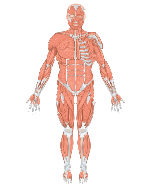
Pick the name of the highlighted marker.
Keep practicing
Now make your own
Upload your prep book, slides or notes, or just tell Bo what you want to learn. You do not need a document to start.
About this diagram
Tap a marker on the muscular system and pick the muscle it sits on. There are twelve markers, from the neck down to the shin, all on muscles you can see from the front. The figure faces you, so the body's right side is on the left of the picture, and on the right half of the picture the chest muscles are cut back to show the ribs.
All 12 labels on this diagram
Every marker on the figure is listed below with what it does. Answers stay collapsed until you open them, so you can quiz yourself as you scroll.
1.Sternocleidomastoid
The strap-like muscle running from the bony bump behind the ear down to the breastbone and collarbone, which is where its name comes from. One side alone tilts the head toward its own shoulder and turns the face to the opposite side, and both sides together bend the neck forward.
2.Deltoid
The rounded cap of the shoulder, named for its triangular shape like the Greek letter delta. Its front fibers help lift the arm forward, its middle fibers raise it out to the side, and its back fibers pull it backward.
3.Pectoralis major
The large fan-shaped muscle of the upper chest, running from the collarbone, breastbone and upper ribs to the upper arm bone. It pulls the arm across the body and rotates it inward, which is the push in a push-up or a bench press.
4.Biceps brachii
The two-headed muscle on the front of the upper arm, which is what biceps means. It bends the elbow and also turns the forearm so the palm faces up, so it works hardest when you lift a heavy bag with your palm toward you.
5.Rectus abdominis
The pair of straight vertical straps down the front of the abdomen. Bands of tendon cut each strap into segments, which is why a well-developed one looks like a six-pack. It bends the trunk forward, as in a curl-up, and holds the abdomen firm.
6.External oblique
The largest and most superficial of the flat muscles on the side of the abdomen. Its fibers run downward and inward, the way your hands slide into front pockets. One side twists the trunk toward the opposite side, and both sides together bend the trunk forward.
7.Sartorius
The longest muscle in the body, a thin strap that runs diagonally from the front of the hip bone across the thigh to the inner side of the knee. It bends and turns the hip and also bends the knee, which together give the cross-legged sitting position. The name comes from the Latin for tailor.
8.Rectus femoris
The straight muscle in the middle of the front of the thigh, and the one quadriceps muscle that crosses the hip as well as the knee. It extends the knee and also helps lift the thigh toward the chest.
9.Vastus lateralis
The largest of the four quadriceps muscles, running down the outer side of the thigh. Vastus means large and lateralis means toward the side. It extends the knee, and its bulge above the knee is easy to spot on a front view.
10.Vastus medialis
The teardrop-shaped quadriceps muscle on the inner side of the thigh, bulging just above the kneecap. Medialis means toward the middle. It extends the knee, and its lowest fibers help keep the kneecap tracking in line.
11.Tibialis anterior
The muscle on the front of the shin, lying just outside the shin bone, which is the tibia. It lifts the front of the foot toward the shin and helps turn the sole inward. When it stops working, the toes drag and the foot slaps down as you walk, which is called foot drop.
12.Gastrocnemius
The two-headed calf muscle. From the front you only see the edge of its inner head beside the shin bone, so the marker sits on that narrow strip. It points the foot downward, as when you rise onto your toes, and with the soleus it forms the Achilles tendon.
How muscles get their names, and why that makes labeling easier
Most muscle names tell you where the muscle is, what it looks like or what it does, so you can often work a name out instead of memorizing it. Six clues cover most of the twelve on this page.
Location: tibialis anterior is the muscle at the front of the tibia, and sternocleidomastoid is named for the three places it attaches, the sternum, the clavicle and the mastoid bone behind the ear. Shape: the deltoid is a triangle. Size: vastus means large. Number of heads: biceps has two, triceps three and quadriceps four. Direction of fibers: rectus means straight and oblique means slanted. Action: flexor bends a joint, extensor straightens it and adductor pulls toward the midline.
Put the clues together and a name like rectus abdominis reads as the straight muscle of the abdomen, and vastus lateralis as the large muscle on the outer side. Femoris means of the thigh, so rectus femoris is the straight muscle of the thigh. When a marker looks unfamiliar, ask which of the six clues the answer list is offering you.
The pairs that get mixed up, and what the figure leaves out
Most lost marks on a muscle diagram come from a few pairs.
Deltoid and pectoralis major. The deltoid is the rounded cap over the shoulder joint. The pectoralis major is the fan across the chest, and its fibers all converge toward the upper arm.
The quadriceps. It is four muscles, not one: rectus femoris in the middle of the thigh, vastus lateralis on the outer side, vastus medialis on the inner side above the kneecap, and vastus intermedius, which lies under the rectus femoris and cannot be seen from the front. Three of the four are marked here.
Tibialis anterior and gastrocnemius. One is the front of the shin and the other is the calf. From the front only a narrow strip of the calf shows beside the shin bone, which is why its marker sits on that strip.
Rectus abdominis and external oblique. The rectus is the straight, segmented strip down the middle, and the external oblique is the broad sheet at the side of it.
Left and right always belong to the body in the picture, not to you. The figure also shows muscles that are not marked, such as the face muscles, the forearm and the foot, and it leaves out the muscles of the back, such as the latissimus dorsi, the gluteus maximus and the hamstrings, which need a view from behind.
How StudyPDF turns your own diagrams into labeling practice
Twelve markers on one figure are a good start. An anatomy practical will use the diagram from your own textbook or slides, often a view from behind or a close up of one limb, with the labels your instructor picked.
StudyPDF works from your material instead. Upload the chapter, the lecture slides or the worksheet. When your slides have a labeled diagram of the muscles, you can turn it into image occlusion flashcards from the New material form: the labels are covered and you recall each one. Quizzes and practice tests come from the same pages, and every explanation cites where the answer came from. Bo, the tutor, explains any muscle you get wrong from your own pages.
You do not need a file to start. Name a topic instead, for example the muscles of the thigh or how muscles are named, and Bo sets it up so you can build practice from it. Starting is free.
Written by the StudyPDF team. Last updated August 19, 2026.
Good to know
Is this muscle labeling quiz free?
Yes. All twelve markers, your score and the explanation for every muscle are free to play in your browser. The printable label list needs a free StudyPDF account.
Which muscles do I need to know for an intro anatomy course?
Courses differ, so check your syllabus. The twelve marked here are the large surface muscles that nearly every intro course starts with. Many courses add the trapezius, latissimus dorsi, gluteus maximus and hamstrings, which are on the back of the body.
What are the four quadriceps muscles?
Rectus femoris, vastus lateralis, vastus medialis and vastus intermedius. Quad means four. The vastus intermedius lies under the rectus femoris, so it cannot be seen on a front view like this one.
Can I print this as a worksheet?
Yes. The download button gives you a numbered list of the twelve muscles with an explanation of each, which works as a revision sheet or as an answer key next to a blank diagram.