Label the Ear
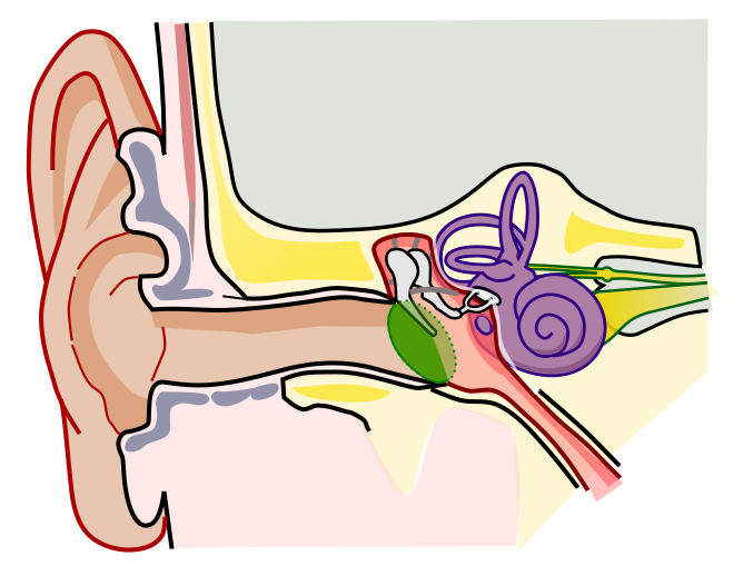
Tap a marker on the ear and pick the part it points to. There are ten, from the auricle on the outside to the nerve that carries sound to the brain. The figure is a cut through the side of the head, with the outer ear on the left and the inner ear on the right, so sound travels from left to right across the picture.
Pick the name of the highlighted marker.
All Nursing & Health quizzes
- Labeling quiz · 12 labels
Label the Eye Diagram
- Labeling quiz · 10 labels
Label the Neuron
- Practice test · 15 questions
Anatomy and Physiology Practice Test

Now make your own
Upload your prep book, slides or notes, or just tell Bo what you want to learn. You do not need a document to start.
All 10 labels on this diagram
Every marker on the figure is listed below with what it does. Answers stay collapsed until you open them, so you can quiz yourself as you scroll.
1. Auricle (pinna)
Show answer
The visible flap on the side of the head, made of cartilage covered in skin, except for the soft earlobe at the bottom. Its folds catch sound and funnel it into the ear canal, and they also change the sound slightly depending on where it comes from, which helps you tell above from below and front from back.
2. External auditory canal
Show answer
The tube from the auricle to the eardrum, about 2.5 centimeters, or an inch, long in an adult. Glands in its outer part make earwax, which traps dust and keeps the skin from drying out. On the figure it is the long tan channel running across the middle.
3. Tympanic membrane
Show answer
The eardrum, a thin membrane stretched across the end of the ear canal that separates the outer ear from the middle ear. Sound waves make it vibrate, and it passes those vibrations on to the malleus, whose handle is attached to its inner side. That handle is the dark stripe running across the green membrane on this figure.
4. Malleus
Show answer
The hammer, the first and largest of the three tiny bones of the middle ear, called the ossicles. Its handle is fixed to the eardrum, so it moves every time the eardrum vibrates, and its head connects to the incus. Here the marker sits on its rounded head.
5. Incus
Show answer
The anvil, the middle one of the three ossicles. Its body is joined to the head of the malleus, and its long process reaches down to meet the stapes, so it relays the movement from one to the other. The marker sits on that long process.
6. Stapes
Show answer
The stirrup, the smallest bone in the human body at roughly 3 millimeters. Its flat footplate sits in the oval window, an opening into the inner ear, and pushes on the fluid inside. Because the eardrum is many times larger than that footplate, the ossicles concentrate the sound's force onto a small area, which is how sound in air can move fluid.
7. Semicircular canals
Show answer
Three fluid-filled loops set at right angles to each other, one for each direction the head can turn. When you turn your head, the fluid lags behind and bends tiny hair cells, which tells the brain about the rotation. They are for balance, not hearing.
8. Cochlea
Show answer
The snail-shaped hearing organ, a fluid-filled tube coiled two and a half to almost three times. Movement of the stapes sends waves through that fluid, and hair cells along its length turn them into nerve signals. Each pitch has its own place: high sounds are picked up near the base and low sounds near the tip.
9. Vestibulocochlear nerve
Show answer
Cranial nerve VIII, which carries signals from the inner ear to the brainstem. It has two parts: the cochlear nerve for hearing, the broad yellow band from the cochlea where this marker sits, and the vestibular nerve for balance, the thin strand just above it with a small swelling.
10. Eustachian tube
Show answer
The narrow tube that runs from the middle ear down to the back of the nose and throat, also called the auditory tube. It opens when you swallow or yawn to let air in or out, which keeps the pressure on both sides of the eardrum equal and is the pop you feel on a plane. In young children it is shorter and more level, one reason middle ear infections are more common at that age.
Outer, middle and inner ear: follow the sound
Most ear diagrams look busy, but every labeled part belongs to one of three sections, and exams usually ask you to sort them as well as name them.
The outer ear catches sound and carries it inward. It is the auricle and the external auditory canal, and it ends at the tympanic membrane, the eardrum.
The middle ear is a small air-filled space behind the eardrum, the deeper pink area on this figure. It holds the three ossicles, the malleus, the incus and the stapes, which pass the vibration along like a chain of levers. The Eustachian tube leaves the middle ear and runs down to the throat, which is why a cold can make your ears feel blocked.
The inner ear is filled with fluid and sits deep in the skull bone, shown in purple. The cochlea handles hearing, the semicircular canals handle balance, and the vestibulocochlear nerve carries both kinds of signal to the brain.
If you can say the path in order, you can place most markers on any ear diagram: auricle, ear canal, eardrum, malleus, incus, stapes, cochlea, nerve. Air becomes vibration in the eardrum, vibration becomes movement of the ossicles, movement becomes waves in fluid, and waves become nerve signals.
The parts that get mixed up, and what is on the figure but not marked
Most lost marks on an ear diagram come from a few spots.
The ossicles. They always run in the same order from the eardrum inward: malleus, incus, stapes, or hammer, anvil, stirrup. The malleus is the one touching the eardrum and the stapes is the one touching the inner ear, so if you know the two ends, the incus is whatever is left in the middle.
Cochlea and semicircular canals. Both are purple on this figure and both belong to the inner ear. The coiled snail shell is the cochlea and is for hearing. The loops above it are the semicircular canals and are for balance.
Eardrum and middle ear. The eardrum is the membrane, the middle ear is the space behind it. A marker on the green oval is the tympanic membrane, never the middle ear.
Several structures are visible but deliberately left unmarked. The oval window is the opening where the footplate of the stapes sits. The round window is the small purple oval just below it, a second membrane that bulges out as the stapes pushes in, so the fluid has somewhere to go. The vestibule is the rounded purple chamber where the semicircular canals meet the cochlea. The pink space around the ossicles is the tympanic cavity, the air-filled middle ear itself. Some courses expect all of these too, so check your own list.
How StudyPDF turns your own diagrams into labeling practice
Ten markers on one figure are a good start. A biology test or an anatomy practical will use the diagram from your own textbook or slides, often drawn from a different angle, with the labels your teacher picked.
StudyPDF works from your material instead. Upload the chapter, the lecture slides or the worksheet. When your slides have a labeled diagram of the ear, you can turn it into image occlusion flashcards from the New material form: the labels are covered and you recall each one. Quizzes and practice tests come from the same pages, and every explanation cites where the answer came from. Bo, the tutor, explains any part you get wrong from your own pages.
You do not need a file to start. Name a topic instead, for example the parts of the ear and their functions, or how the ear turns sound into nerve signals, and Bo sets it up so you can build practice from it. Starting is free.
Written by the StudyPDF team. Last updated 2026-08-19.