Label the Digestive System
Tap a marker on the diagram and pick the part it belongs to. There are eleven markers, following food from the salivary glands and tongue down through the esophagus, stomach and intestines to the rectum, with the liver, gallbladder and pancreas along the way. The head is cut open from the side and the torso is drawn from the front, so the liver is on the left of the picture.
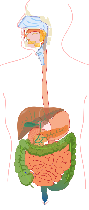
Pick the name of the highlighted marker.
All Nursing & Health quizzes
- Labeling quiz · 11 labels
Label the Respiratory System
- Labeling quiz · 10 labels
Label the Heart Diagram
- Practice test · 15 questions
Anatomy and Physiology Practice Test

Now make your own
Upload your prep book, slides or notes, or just tell Bo what you want to learn. You do not need a document to start.
All 11 labels on this diagram
Every marker on the figure is listed below with what it does. Answers stay collapsed until you open them, so you can quiz yourself as you scroll.
1. Salivary glands
Show answer
Three pairs of glands that make saliva: the parotid, submandibular and sublingual glands. The marker is on the yellow gland at the back of the mouth. Saliva moistens food and starts breaking down starch with the enzyme amylase.
2. Tongue
Show answer
A muscle that moves food around the mouth, pushes it against the teeth and shapes it into a ball called a bolus. It also carries the taste buds. When you swallow, it pushes the bolus back toward the throat.
3. Pharynx
Show answer
The throat, a muscular tube behind the mouth and nose. Air and food both pass through it, and swallowing sends the food on into the esophagus while a flap called the epiglottis keeps it out of the airway.
4. Esophagus
Show answer
The muscular tube that carries food from the throat to the stomach. It does not digest anything. Waves of muscle contraction called peristalsis squeeze the food down, which is why you can swallow even when you are lying down.
5. Liver
Show answer
The largest solid organ in the abdomen, on the right side of the body and so on the left of this picture. It makes bile, which helps digest fat, and it processes the nutrients that come in from the intestines. It also stores some of them, such as glucose in the form of glycogen.
6. Gallbladder
Show answer
A small pouch tucked under the liver. It stores and concentrates the bile the liver makes and squeezes it into the small intestine when a fatty meal arrives. You can live without it, because the liver keeps making bile.
7. Stomach
Show answer
A stretchy muscular bag that churns food with acid and the enzyme pepsin until it becomes a thick liquid called chyme. It empties a little at a time into the small intestine through a ring of muscle at its lower end.
8. Pancreas
Show answer
The orange band behind the stomach, drawn with its branching ducts. It makes digestive enzymes for starch, protein and fat and sends them into the small intestine, along with bicarbonate that neutralizes stomach acid. The same organ also makes the hormones insulin and glucagon.
9. Small intestine
Show answer
The long, coiled tube where most digestion finishes and nearly all nutrients are absorbed into the blood. Its lining is folded into tiny fingers called villi, which give it a huge surface area. It has three parts: the duodenum, jejunum and ileum.
10. Large intestine
Show answer
The wider tube, shown in green, that frames the small intestine on three sides. It absorbs water and salts from what is left and turns it into stool. The marker is on the ascending colon, which rises on the right side of the body; the transverse colon crosses the top and the descending colon runs down the other side.
11. Rectum
Show answer
The last section of the large intestine, the bluish bulb at the bottom. It holds stool until you are ready to pass it, and stretching of its wall is what creates the urge. The opening to the outside below it is the anus.
Follow one bite and the labels order themselves
The digestive system is easiest to label as a route, because food only moves one way and nearly every marker sits on that path.
It starts in the mouth, where the teeth and tongue break food up and the salivary glands wet it. The swallow carries it through the pharynx and down the esophagus into the stomach. From the stomach it enters the small intestine, then the large intestine, and the waste leaves through the rectum and anus.
That sentence covers eight of the eleven markers in order. The other three are the liver, the gallbladder and the pancreas, which food never enters. They are accessory organs: they make or store the bile and enzymes that are poured into the small intestine so that digestion can finish there.
Reading this figure: the drawing is flattened
Two things in the picture are easy to misread. First, sides. The torso faces you, so the liver sits on the left of the picture even though it belongs to the right side of the body. The stomach is on the right of the picture.
Second, overlap. Real organs sit in layers, and the drawing shows them semi transparent. The pancreas is the orange band behind the stomach, and the green branching lines are the ducts that carry bile and pancreatic juice. The transverse colon, the green band across the middle, passes in front of the small intestine. The small dotted arrow at the lower left of the picture, where the small intestine meets the large, is part of the artwork and not a marker.
It also helps to learn the large intestine as a frame. It rises on one side, crosses the top and drops down the other, which is how the three colon labels earn their names: ascending, transverse and descending.
How StudyPDF turns your own diagrams into labeling practice
One figure with eleven markers will get you through a quiz. An anatomy exam or a lab practical will use the diagrams from your own course, drawn from a different angle, with the terms your instructor expects.
StudyPDF works from your material instead. Upload the chapter, the lecture slides or your lab manual, and Bo, the study agent, builds practice from those exact pages, including the figures in them. Every explanation cites where the answer came from, so you can go straight back to the page.
You do not need a file to start. Name a topic instead, for example the stages of digestion or the role of bile, and Bo sets up practice on it. Starting is free.
Written by the StudyPDF team. Last updated 2026-08-19.