Etiqueta el esqueleto
Las etiquetas se quedan en inglés.
Toca un marcador del esqueleto y elige el hueso al que corresponde. Hay doce marcadores, que van desde el cráneo hasta la espinilla. La figura está de frente, así que el lado derecho del esqueleto queda a la izquierda de la imagen, y aquí todos los marcadores de brazos y piernas están en sus extremidades izquierdas.
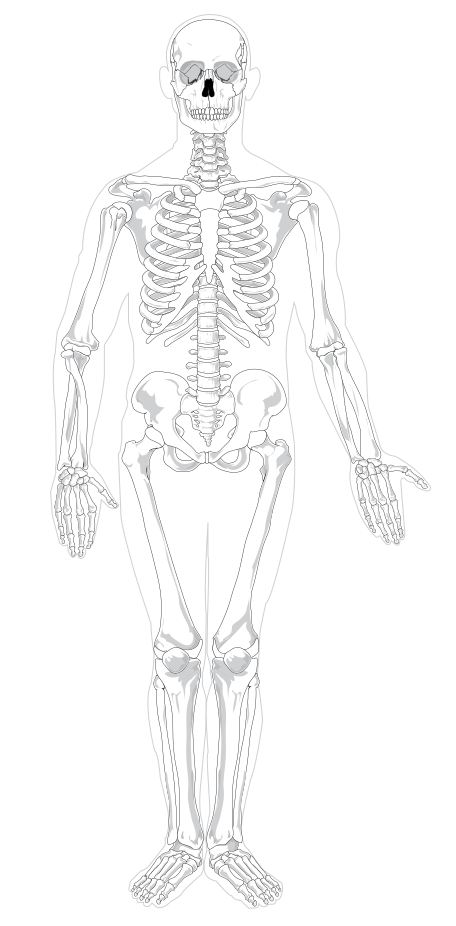
Elige el nombre del punto marcado.
Todos los tests de Enfermería y salud
- Cuestionario de etiquetado · 10 etiquetas
Etiqueta el diagrama del corazón
- Test de práctica · 15 preguntas
Test de práctica de anatomía y fisiología
- Cuestionario de etiquetado · 10 etiquetas
Etiqueta la neurona

Crea el tuyo
Sube tu libro de preparación, tus diapositivas o tus apuntes, o dile a Bo qué quieres estudiar. No necesitas ningún documento para empezar.
Las 12 etiquetas de este diagrama
Abajo están todos los marcadores de la figura con su función. Las respuestas quedan plegadas para que puedas autoevaluarte mientras bajas.
1. Skull
Ver respuesta
The bony case around the brain, made of eight cranial bones fused at jagged seams called sutures. Everything above the eye sockets on this figure is cranium.
2. Mandible
Ver respuesta
The lower jaw, and the only bone in the skull that moves. It carries the lower teeth and hinges at the temporomandibular joint just in front of each ear.
3. Clavicle
Ver respuesta
The collarbone, running from the top of the sternum out to the shoulder. It props the shoulder away from the chest, and its shallow S shape is easy to spot on any front view.
4. Sternum
Ver respuesta
The breastbone down the middle of the chest. The upper ribs join it through costal cartilage, which is why the rib cage can spring outwards when you breathe in.
5. Rib
Ver respuesta
One of the twelve pairs that form the cage protecting the heart and lungs. The top seven pairs reach the sternum directly, three join the cartilage above them, and the bottom two are the floating ribs.
6. Humerus
Ver respuesta
The single bone of the upper arm, from the shoulder joint to the elbow. It is the long bone people mean when they say they have broken an arm above the elbow.
7. Ulna
Ver respuesta
The forearm bone on the little finger side. It is the thicker of the two at the elbow, where its hooked end forms the point you can feel when you bend your arm.
8. Radius
Ver respuesta
The forearm bone on the thumb side. It is thin at the elbow and broad at the wrist, and it is the one that rotates over the ulna when you turn your palm downwards.
9. Pelvis
Ver respuesta
The basin of bone carrying the weight of the upper body into the legs. Each side is three bones fused together, and the broad flared blade you can see here is the ilium.
10. Femur
Ver respuesta
The thigh bone, the longest and strongest bone in the body. Its ball shaped head sits in the socket of the pelvis, and it angles inwards so the knees end up under the body's centre of gravity.
11. Patella
Ver respuesta
The kneecap, a sesamoid bone sitting inside the tendon of the thigh muscles. It is not joined to the leg bones by a joint of its own, it rides over the front of the knee as it bends.
12. Tibia
Ver respuesta
The shin bone, the thicker and inner of the two bones in the lower leg. It takes almost all the weight, which is why a broken tibia stops you walking and a broken fibula often does not.
Axial o apendicular, y por qué conviene saber la división
Un esqueleto adulto tiene unos 206 huesos, y ningún examen te pide nombrarlos todos. Lo que sí piden los exámenes es la división en dos grupos, porque ordena todo lo demás.
El esqueleto axial es el núcleo: el cráneo, la columna vertebral, las costillas y el esternón. Protege el encéfalo, la médula espinal, el corazón y los pulmones, y apenas se mueve. En esta figura es la cabeza, la columna que baja por el centro y toda la caja torácica.
El esqueleto apendicular son las extremidades y las cinturas de las que cuelgan: la clavícula y la escápula en el hombro, el brazo y la mano, la pelvis, y la pierna y el pie. Está hecho para el movimiento y no para la protección. De los doce marcadores de aquí, cinco son axiales y siete apendiculares.
La cifra en sí merece un matiz. Los bebés nacen con unos 270 huesos y muchos se fusionan durante el crecimiento, y los adultos varían de verdad, así que 206 es la cifra habitual más que una cifra fija.
Las parejas que se confunden, y cómo distinguirlas
Casi todos los puntos que se pierden en un diagrama del esqueleto se van en una de tres confusiones.
Radio y cúbito. El radio está del lado del pulgar, fino en el codo y ancho en la muñeca. El cúbito está del lado del meñique, grueso en el codo y fino en la muñeca. La regla que casi todo el mundo se queda es que el radio rota, que es lo que te permite girar la palma.
Tibia y peroné. La tibia es el hueso interno y grueso que carga el peso, y el peroné el fino de fuera, que sobre todo sirve de anclaje a los músculos y forma el tobillo por su cara externa. El peroné se ve en esta figura como el hueso delgado que hay junto a la tibia, pero a propósito no está marcado, porque a esta escala un marcador suyo quedaría casi encima del de la tibia.
Izquierda y derecha. La figura está de frente, así que su lado derecho queda a tu izquierda. La izquierda y la derecha anatómicas son siempre las del cuerpo que se describe, nunca las de quien mira.
Dos estructuras más que vale la pena buscar mientras estás aquí, ninguna de las dos marcada: la escápula, el omóplato, que queda casi entera por detrás de la caja torácica y solo se ve en parte desde delante, y la columna vertebral, que baja por el centro y puedes ver entre las costillas y por encima de la pelvis.
Cómo StudyPDF convierte tus propios diagramas en práctica de etiquetado
Doce marcadores en una figura te sacan de un cuestionario. No te sacan de un examen práctico de laboratorio, donde el diagrama es el de tu propio libro, muchas veces una vista posterior o un primer plano de una sola región, y con las etiquetas que usó tu profesor.
StudyPDF parte de tu material. Sube el capítulo, las diapositivas de clase o el manual de laboratorio, y Bo, el agente de estudio, crea práctica a partir de esas páginas exactas, incluidas las figuras que contienen. Cada explicación cita de dónde salió la respuesta, así que puedes volver a la página en cuanto algo te chirríe.
No necesitas un archivo para empezar. Nombra un tema, por ejemplo los huesos del esqueleto axial o los huesos de la mano, y Bo crea práctica con eso. Empezar es gratis.
Escrito por el equipo de StudyPDF. Actualizado el 2026-08-19.