Etiqueta los huesos del cráneo
Las etiquetas se quedan en inglés.
Toca un marcador del cráneo y elige el hueso al que pertenece. Hay once, uno por cada hueso que aparece en su propio color, desde el hueso frontal de la frente hasta la mandíbula. El cráneo mira hacia la izquierda, así que ves su lado izquierdo, y los marcadores 6 y 7 están muy juntos dentro de la órbita del ojo.
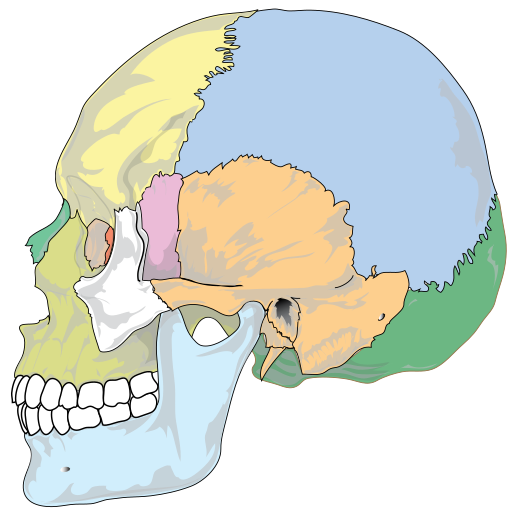
Elige el nombre del punto marcado.
Todos los tests de Enfermería y salud
- Cuestionario de etiquetado · 12 etiquetas
Etiqueta el esqueleto
- Cuestionario de etiquetado · 12 etiquetas
Etiqueta el diagrama del ojo
- Test de práctica · 15 preguntas
Test de práctica de anatomía y fisiología

Crea el tuyo
Sube tu libro de preparación, tus diapositivas o tus apuntes, o dile a Bo qué quieres estudiar. No necesitas ningún documento para empezar.
Las 11 etiquetas de este diagrama
Abajo están todos los marcadores de la figura con su función. Las respuestas quedan plegadas para que puedas autoevaluarte mientras bajas.
1. Frontal bone
Ver respuesta
The bone of the forehead, which also forms the roof of each eye socket. It meets the two parietal bones at the coronal suture, the wavy seam running over the top of the head behind it.
2. Parietal bone
Ver respuesta
One of a pair that makes up most of the roof and the upper sides of the skull. The two parietal bones meet each other along the sagittal suture on top of the head, which this side view cannot show.
3. Temporal bone
Ver respuesta
The bone at the side of the head, below the parietal. It houses the middle and inner ear, and the dark opening just behind the jaw joint on this figure is the ear canal, the external acoustic meatus. The mandible hinges against it at the temporomandibular joint.
4. Occipital bone
Ver respuesta
The back and base of the skull. The spinal cord passes through its large opening, the foramen magnum, and the two occipital condyles beside that opening rest on the first vertebra, the atlas.
5. Sphenoid bone
Ver respuesta
A single bat-shaped bone that runs right across the skull base. From the side you only see its greater wing at the temple, but it touches every other cranial bone, and a hollow on its upper surface, the sella turcica, holds the pituitary gland.
6. Ethmoid bone
Ver respuesta
A light, spongy bone between the eye sockets that forms the roof and much of the upper walls of the nasal cavity. From the side you only see a thin plate of it in the inner wall of the eye socket, the narrow red strip under this marker. The nerves for smell pass through its cribriform plate.
7. Lacrimal bone
Ver respuesta
The smallest bone of the face, a thin plate in the inner wall of the eye socket, in front of the ethmoid. A groove in it holds the lacrimal sac, which drains tears down into the nose.
8. Nasal bone
Ver respuesta
One of two small bones side by side that form the bony bridge of the nose. Only the upper part of the nose is bone. The rest is cartilage, which is why a skull has an open hole where the tip of the nose would be.
9. Zygomatic bone
Ver respuesta
The cheekbone, forming the outer wall and part of the floor of the eye socket. Its back end joins a projection of the temporal bone, and the two together make the zygomatic arch you can feel in front of your ear.
10. Maxilla
Ver respuesta
The upper jaw, which carries the upper teeth. The two maxillae meet in the middle, form most of the hard palate and the floor of each eye socket, and each one holds the largest of the air-filled sinuses, the maxillary sinus.
11. Mandible
Ver respuesta
The lower jaw, the largest and strongest bone of the face and the only skull bone that moves. Its upright back part, the ramus, rises to the joint with the temporal bone just in front of the ear canal.
Veintidós huesos, y los once que se ven de lado
Un cráneo adulto se suele contar como 22 huesos, divididos en dos grupos. Los 8 huesos craneales forman la caja que rodea el cerebro: un frontal, dos parietales, dos temporales, un occipital, un esfenoides y un etmoides. Los 14 huesos faciales forman la cara: dos nasales, dos maxilares, dos cigomáticos, dos lagrimales, dos palatinos, dos cornetes nasales inferiores, un vómer y una mandíbula.
Una vista lateral muestra once de esos nombres, y son los once marcadores de aquí. Seis son craneales: frontal, parietal, temporal, occipital, esfenoides y etmoides. Cinco son faciales: lagrimal, nasal, cigomático, maxilar y mandíbula. Los huesos pares solo aparecen una vez, porque el otro está en el lado opuesto del cráneo.
Los palatinos, el vómer y los cornetes nasales inferiores faltan por una razón sencilla: están en lo profundo de la nariz y la boca y no se ven de lado. Los huesecillos del oído y el hioides del cuello tampoco cuentan entre los 22, y por eso algunos libros dan totales más altos.
Suturas, puntos de referencia y las confusiones que quitan puntos
Los huesos craneales están unidos por suturas, costuras dentadas que son flexibles en un bebé y se van fusionando con la edad. De lado se ven tres. La sutura coronal va entre el frontal y los parietales, la lambdoidea entre los parietales y el occipital, y la escamosa se curva por el borde superior del temporal. La cuarta grande, la sutura sagital entre los dos parietales, recorre la parte de arriba de la cabeza y solo se ve desde arriba.
Hay un punto de referencia que sale en casi todos los exámenes: el pterion, la pequeña zona de la sien donde se unen el frontal, el parietal, el temporal y el esfenoides. Es una de las partes más finas de la pared del cráneo, y justo por debajo pasa la arteria meníngea media. En esta figura está más o menos en la parte de arriba del esfenoides rosa.
Los errores en este diagrama son previsibles. El esfenoides y el temporal se intercambian porque los dos se ven en el lado de la cabeza: el esfenoides es la franja estrecha de delante y el temporal la placa ancha de detrás. El arco cigomático son dos huesos, no uno, el cigomático delante y una prolongación del temporal detrás. En la órbita, el lagrimal es la lámina de delante y el etmoides está detrás. Y el maxilar es el hueso de la arcada superior, fijo al cráneo, mientras que la mandíbula es el de la arcada inferior, el que se mueve.
Cómo StudyPDF convierte tus propios diagramas en práctica de etiquetado
Once marcadores en una figura te bastan para un cuestionario. Un práctico de laboratorio es más difícil: el cráneo suele ser uno real o la figura de tu propio libro, puede ser una vista frontal o la base del cráneo, y el profesor puede pedirte agujeros y apófisis además de los huesos enteros.
StudyPDF parte de tu material. Sube el capítulo, las diapositivas de clase o el manual de laboratorio, y Bo, el agente de estudio, crea práctica a partir de esas páginas exactas, incluidas sus figuras. Cada explicación cita de dónde salió la respuesta, así que puedes volver directo a la página cuando algo no te cuadre.
No necesitas un archivo para empezar. Nombra un tema, por ejemplo los huesos craneales y las suturas o los agujeros de la base del cráneo, y Bo crea práctica a partir de eso. Empezar es gratis.
Escrito por el equipo de StudyPDF. Actualizado el 2026-08-19.