Etiqueta las partes del oído
Las etiquetas se quedan en inglés.
Toca un marcador del oído y elige la parte a la que apunta. Hay diez, desde el pabellón auricular por fuera hasta el nervio que lleva el sonido al cerebro. La figura es un corte por el lado de la cabeza, con el oído externo a la izquierda y el oído interno a la derecha, así que el sonido viaja de izquierda a derecha por la imagen.
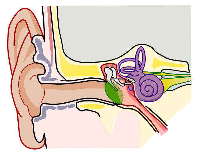
Elige el nombre del punto marcado.
Todos los tests de Enfermería y salud
- Cuestionario de etiquetado · 12 etiquetas
Etiqueta el diagrama del ojo
- Cuestionario de etiquetado · 10 etiquetas
Etiqueta la neurona
- Test de práctica · 15 preguntas
Test de práctica de anatomía y fisiología

Crea el tuyo
Sube tu libro de preparación, tus diapositivas o tus apuntes, o dile a Bo qué quieres estudiar. No necesitas ningún documento para empezar.
Las 10 etiquetas de este diagrama
Abajo están todos los marcadores de la figura con su función. Las respuestas quedan plegadas para que puedas autoevaluarte mientras bajas.
1. Auricle (pinna)
Ver respuesta
The visible flap on the side of the head, made of cartilage covered in skin, except for the soft earlobe at the bottom. Its folds catch sound and funnel it into the ear canal, and they also change the sound slightly depending on where it comes from, which helps you tell above from below and front from back.
2. External auditory canal
Ver respuesta
The tube from the auricle to the eardrum, about 2.5 centimeters, or an inch, long in an adult. Glands in its outer part make earwax, which traps dust and keeps the skin from drying out. On the figure it is the long tan channel running across the middle.
3. Tympanic membrane
Ver respuesta
The eardrum, a thin membrane stretched across the end of the ear canal that separates the outer ear from the middle ear. Sound waves make it vibrate, and it passes those vibrations on to the malleus, whose handle is attached to its inner side. That handle is the dark stripe running across the green membrane on this figure.
4. Malleus
Ver respuesta
The hammer, the first and largest of the three tiny bones of the middle ear, called the ossicles. Its handle is fixed to the eardrum, so it moves every time the eardrum vibrates, and its head connects to the incus. Here the marker sits on its rounded head.
5. Incus
Ver respuesta
The anvil, the middle one of the three ossicles. Its body is joined to the head of the malleus, and its long process reaches down to meet the stapes, so it relays the movement from one to the other. The marker sits on that long process.
6. Stapes
Ver respuesta
The stirrup, the smallest bone in the human body at roughly 3 millimeters. Its flat footplate sits in the oval window, an opening into the inner ear, and pushes on the fluid inside. Because the eardrum is many times larger than that footplate, the ossicles concentrate the sound's force onto a small area, which is how sound in air can move fluid.
7. Semicircular canals
Ver respuesta
Three fluid-filled loops set at right angles to each other, one for each direction the head can turn. When you turn your head, the fluid lags behind and bends tiny hair cells, which tells the brain about the rotation. They are for balance, not hearing.
8. Cochlea
Ver respuesta
The snail-shaped hearing organ, a fluid-filled tube coiled two and a half to almost three times. Movement of the stapes sends waves through that fluid, and hair cells along its length turn them into nerve signals. Each pitch has its own place: high sounds are picked up near the base and low sounds near the tip.
9. Vestibulocochlear nerve
Ver respuesta
Cranial nerve VIII, which carries signals from the inner ear to the brainstem. It has two parts: the cochlear nerve for hearing, the broad yellow band from the cochlea where this marker sits, and the vestibular nerve for balance, the thin strand just above it with a small swelling.
10. Eustachian tube
Ver respuesta
The narrow tube that runs from the middle ear down to the back of the nose and throat, also called the auditory tube. It opens when you swallow or yawn to let air in or out, which keeps the pressure on both sides of the eardrum equal and is the pop you feel on a plane. In young children it is shorter and more level, one reason middle ear infections are more common at that age.
Oído externo, medio e interno: sigue el sonido
La mayoría de diagramas del oído parecen recargados, pero cada parte etiquetada pertenece a una de tres secciones, y los exámenes suelen pedirte que las clasifiques además de nombrarlas.
El oído externo capta el sonido y lo lleva hacia dentro. Lo forman el pabellón auricular y el conducto auditivo externo, y termina en la membrana timpánica, el tímpano.
El oído medio es un pequeño espacio lleno de aire detrás del tímpano, la zona rosa más oscura de esta figura. Contiene los tres huesecillos, el martillo, el yunque y el estribo, que transmiten la vibración como una cadena de palancas. La trompa de Eustaquio sale del oído medio y baja hasta la garganta, y por eso con un resfriado puedes notar los oídos tapados.
El oído interno está lleno de líquido y se aloja en lo profundo del hueso del cráneo, en morado en la figura. La cóclea se encarga de la audición, los conductos semicirculares del equilibrio, y el nervio vestibulococlear lleva los dos tipos de señal al cerebro.
Si sabes decir el recorrido en orden, puedes situar la mayoría de los marcadores en cualquier diagrama del oído: pabellón auricular, conducto auditivo, tímpano, martillo, yunque, estribo, cóclea, nervio. El aire se convierte en vibración en el tímpano, la vibración en movimiento de los huesecillos, el movimiento en ondas en un líquido, y las ondas en señales nerviosas.
Las partes que se confunden y lo que sale en la figura sin marcar
La mayoría de puntos perdidos en un diagrama del oído vienen de unos pocos sitios.
Los huesecillos. Siempre van en el mismo orden desde el tímpano hacia dentro: martillo, yunque y estribo, o en latín malleus, incus y stapes. El martillo es el que toca el tímpano y el estribo el que toca el oído interno, así que si te sabes los dos extremos, el yunque es el que queda en medio.
Cóclea y conductos semicirculares. Los dos son morados en esta figura y los dos pertenecen al oído interno. La concha de caracol enrollada es la cóclea y sirve para oír. Los bucles de encima son los conductos semicirculares y sirven para el equilibrio.
Tímpano y oído medio. El tímpano es la membrana y el oído medio es el espacio que hay detrás. Un marcador sobre el óvalo verde es la membrana timpánica, nunca el oído medio.
Varias estructuras se ven pero se han dejado sin marcar a propósito. La ventana oval es la abertura donde se apoya la base del estribo. La ventana redonda es el pequeño óvalo morado justo debajo, una segunda membrana que se abomba hacia fuera cuando el estribo empuja hacia dentro, para que el líquido tenga adónde ir. El vestíbulo es la cámara morada redondeada donde se unen los conductos semicirculares y la cóclea. El espacio rosa alrededor de los huesecillos es la cavidad timpánica, el propio oído medio lleno de aire. Algunos cursos piden también todas estas, así que revisa tu propia lista.
Cómo StudyPDF convierte tus propios diagramas en práctica de etiquetado
Diez marcadores en una figura son un buen comienzo. Un examen de biología o un práctico de anatomía usará el diagrama de tu propio libro o de tus diapositivas, a menudo dibujado desde otro ángulo, con las etiquetas que eligió tu profesor.
StudyPDF parte de tu material. Sube el capítulo, las diapositivas de clase o la ficha. Si tus diapositivas tienen un diagrama del oído con etiquetas, puedes convertirlo en tarjetas de oclusión de imagen desde el formulario de Nuevo material: las etiquetas quedan tapadas y tú recuerdas cada una. Los cuestionarios y los tests de práctica salen de esas mismas páginas, y cada explicación cita de dónde salió la respuesta. Bo, el tutor, te explica desde tus propias páginas cualquier parte que falles.
No necesitas un archivo para empezar. Nombra un tema, por ejemplo las partes del oído y sus funciones o cómo convierte el oído el sonido en señales nerviosas, y Bo lo prepara para que puedas crear práctica a partir de él. Empezar es gratis.
Escrito por el equipo de StudyPDF. Actualizado el 2026-08-19.