Etiqueta el aparato digestivo
Las etiquetas se quedan en inglés.
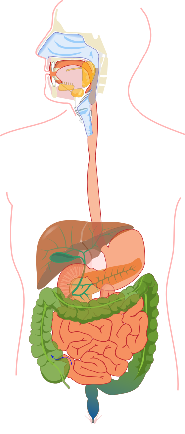
Toca un marcador del diagrama y elige la parte a la que corresponde. Hay once marcadores que siguen el alimento desde las glándulas salivales y la lengua, por el esófago, el estómago y los intestinos, hasta el recto, con el hígado, la vesícula biliar y el páncreas por el camino. La cabeza está cortada de lado y el torso está dibujado de frente, así que el hígado queda a la izquierda de la imagen.
Elige el nombre del punto marcado.
Todos los tests de Enfermería y salud
- Cuestionario de etiquetado · 11 etiquetas
Etiqueta el sistema respiratorio
- Cuestionario de etiquetado · 10 etiquetas
Etiqueta el diagrama del corazón
- Test de práctica · 15 preguntas
Test de práctica de anatomía y fisiología

Crea el tuyo
Sube tu libro de preparación, tus diapositivas o tus apuntes, o dile a Bo qué quieres estudiar. No necesitas ningún documento para empezar.
Las 11 etiquetas de este diagrama
Abajo están todos los marcadores de la figura con su función. Las respuestas quedan plegadas para que puedas autoevaluarte mientras bajas.
1. Salivary glands
Ver respuesta
Three pairs of glands that make saliva: the parotid, submandibular and sublingual glands. The marker is on the yellow gland at the back of the mouth. Saliva moistens food and starts breaking down starch with the enzyme amylase.
2. Tongue
Ver respuesta
A muscle that moves food around the mouth, pushes it against the teeth and shapes it into a ball called a bolus. It also carries the taste buds. When you swallow, it pushes the bolus back toward the throat.
3. Pharynx
Ver respuesta
The throat, a muscular tube behind the mouth and nose. Air and food both pass through it, and swallowing sends the food on into the esophagus while a flap called the epiglottis keeps it out of the airway.
4. Esophagus
Ver respuesta
The muscular tube that carries food from the throat to the stomach. It does not digest anything. Waves of muscle contraction called peristalsis squeeze the food down, which is why you can swallow even when you are lying down.
5. Liver
Ver respuesta
The largest solid organ in the abdomen, on the right side of the body and so on the left of this picture. It makes bile, which helps digest fat, and it processes the nutrients that come in from the intestines. It also stores some of them, such as glucose in the form of glycogen.
6. Gallbladder
Ver respuesta
A small pouch tucked under the liver. It stores and concentrates the bile the liver makes and squeezes it into the small intestine when a fatty meal arrives. You can live without it, because the liver keeps making bile.
7. Stomach
Ver respuesta
A stretchy muscular bag that churns food with acid and the enzyme pepsin until it becomes a thick liquid called chyme. It empties a little at a time into the small intestine through a ring of muscle at its lower end.
8. Pancreas
Ver respuesta
The orange band behind the stomach, drawn with its branching ducts. It makes digestive enzymes for starch, protein and fat and sends them into the small intestine, along with bicarbonate that neutralizes stomach acid. The same organ also makes the hormones insulin and glucagon.
9. Small intestine
Ver respuesta
The long, coiled tube where most digestion finishes and nearly all nutrients are absorbed into the blood. Its lining is folded into tiny fingers called villi, which give it a huge surface area. It has three parts: the duodenum, jejunum and ileum.
10. Large intestine
Ver respuesta
The wider tube, shown in green, that frames the small intestine on three sides. It absorbs water and salts from what is left and turns it into stool. The marker is on the ascending colon, which rises on the right side of the body; the transverse colon crosses the top and the descending colon runs down the other side.
11. Rectum
Ver respuesta
The last section of the large intestine, the bluish bulb at the bottom. It holds stool until you are ready to pass it, and stretching of its wall is what creates the urge. The opening to the outside below it is the anus.
Sigue un bocado y las etiquetas se ordenan solas
El aparato digestivo es más fácil de etiquetar como un recorrido, porque el alimento solo avanza en un sentido y casi todos los marcadores están en ese camino.
Empieza en la boca, donde los dientes y la lengua trituran el alimento y las glándulas salivales lo humedecen. Al tragar, pasa por la faringe y baja por el esófago hasta el estómago. Del estómago entra en el intestino delgado, luego en el intestino grueso, y los desechos salen por el recto y el ano.
Esa frase cubre ocho de los once marcadores, en orden. Los otros tres son el hígado, la vesícula biliar y el páncreas, por donde el alimento nunca pasa. Son órganos accesorios: fabrican o almacenan la bilis y las enzimas que se vierten en el intestino delgado para que la digestión termine allí.
Cómo leer esta figura: el dibujo está aplanado
Hay dos cosas de la imagen que se leen mal con facilidad. La primera, los lados. El torso te mira de frente, así que el hígado queda a la izquierda de la imagen aunque pertenece al lado derecho del cuerpo. El estómago está a la derecha de la imagen.
La segunda, la superposición. Los órganos reales están en capas, y el dibujo los muestra semitransparentes. El páncreas es la banda naranja detrás del estómago, y las líneas verdes ramificadas son los conductos que llevan la bilis y el jugo pancreático. El colon transverso, la banda verde que cruza por el medio, pasa por delante del intestino delgado. La pequeña flecha de puntos en la parte inferior izquierda de la imagen, donde el intestino delgado se une al grueso, forma parte del dibujo y no es un marcador.
También ayuda aprender el intestino grueso como un marco. Sube por un lado, cruza por arriba y baja por el otro, y así se ganan su nombre las tres etiquetas del colon: ascendente, transverso y descendente.
Cómo convierte StudyPDF tus propios diagramas en práctica de etiquetado
Una figura con once marcadores te sirve para un cuestionario. Un examen de anatomía o una práctica de laboratorio usará los diagramas de tu propio curso, dibujados desde otro ángulo, con los términos que espera tu profesor.
StudyPDF parte de tu material. Sube el capítulo, las diapositivas de la clase o tu manual de laboratorio, y Bo, el agente de estudio, crea práctica a partir de esas páginas exactas, incluidas las figuras que traen. Cada explicación cita de dónde sale la respuesta, así que puedes volver directo a la página.
No hace falta un archivo para empezar. Nombra un tema, por ejemplo las etapas de la digestión o la función de la bilis, y Bo prepara práctica sobre él. Empezar es gratis.
Escrito por el equipo de StudyPDF. Actualizado el 2026-08-19.