Identifique as partes do olho
As legendas ficam em inglês.
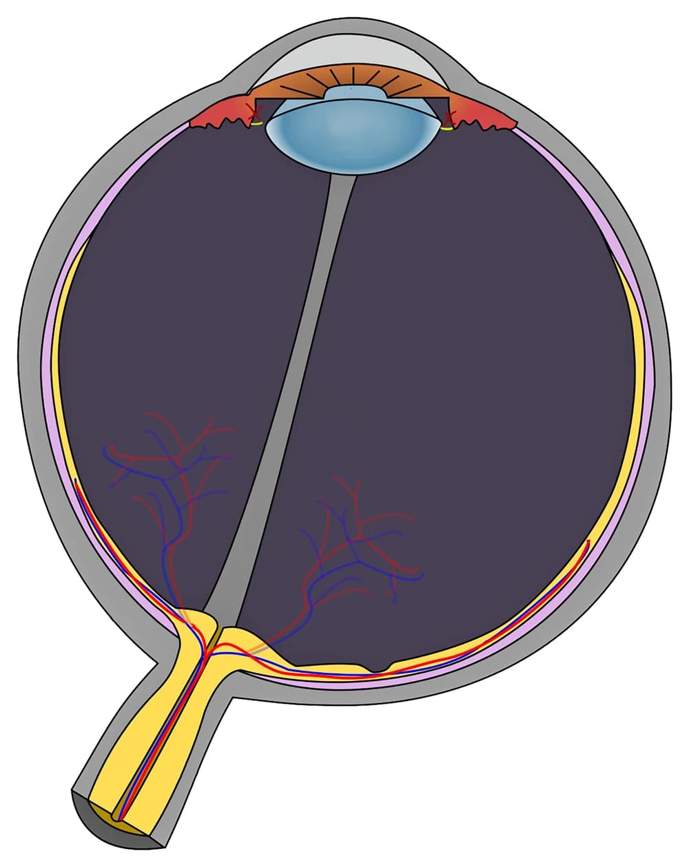
Toque em um marcador do olho e escolha a parte para a qual ele aponta. São doze, da córnea na frente até o nervo óptico atrás. A figura mostra o olho cortado ao meio na horizontal, então a frente do olho fica no alto da imagem e a luz desce através dele até a retina.
Escolhe o nome do marcador destacado.
Todos os quizzes de Enfermagem e Saúde
- Quiz de identificação · 10 identificações
Identifique o neurônio
- Quiz de identificação · 10 identificações
Identifique o diagrama do coração
- Simulado · 15 questões
Simulado de Anatomia e Fisiologia

Crie o seu
Envie seu livro, seus slides ou suas anotações, ou só diga ao Bo o que você quer estudar. Você não precisa de nenhum documento para começar.
Todas as 12 identificações deste diagrama
Abaixo estão todos os marcadores da figura e o que cada um faz. As respostas ficam recolhidas para você se testar enquanto rola a página.
1. Cornea
Mostrar resposta
The clear dome at the very front of the eye. It does most of the focusing, about 70 percent according to the American Academy of Ophthalmology, and the lens only fine tunes what the cornea has already bent. On this figure it is the curved band across the top, where the gray outer wall turns transparent.
2. Aqueous humor
Mostrar resposta
The watery fluid filling the front of the eye between the cornea and the lens, most of it in the anterior chamber between the cornea and the iris. The ciliary body makes it all the time and it drains away at the angle where the iris meets the cornea, and that balance is what keeps the pressure inside the eye steady.
3. Iris
Mostrar resposta
The colored ring you see when you look at someone's eye. It is a muscle: one set of fibers narrows the pupil in bright light and another widens it in the dark. In a cross-section it shows up as the two brown flaps lying in front of the lens.
4. Pupil
Mostrar resposta
The opening in the middle of the iris. It is not a structure at all, just a hole, which is why it looks black from the outside. Here it is the gap between the two halves of the iris, where the front of the lens shows through.
5. Lens
Mostrar resposta
The clear, flexible disc behind the pupil. Thin threads called zonules hold it to the ciliary body, and when the ciliary muscle contracts, those threads slacken and the lens rounds up to focus on something close. It stiffens with age, which is why reading glasses often arrive in your forties.
6. Ciliary body
Mostrar resposta
The red ring of tissue just behind the iris, on both sides of the lens. It does two jobs: its muscle changes the shape of the lens for near and far focus, and its lining produces the aqueous humor.
7. Sclera
Mostrar resposta
The tough outer wall of the eyeball, the part you see as the white of the eye. It keeps the eye's shape and gives the six muscles that move the eye something to pull on. At the front it continues into the cornea.
8. Choroid
Mostrar resposta
The thin layer of blood vessels between the sclera and the retina, shown here in pink. It feeds the outer layers of the retina, and its dark pigment soaks up stray light so it does not bounce around inside the eye.
9. Retina
Mostrar resposta
The light sensitive lining at the back of the eye, shown in yellow. Its rods handle dim light and its cones handle color and fine detail, and together they turn light into nerve signals. On the figure it is the innermost of the three layers in the wall.
10. Vitreous body
Mostrar resposta
The clear gel filling the big chamber between the lens and the retina, which is most of the eyeball. It holds the retina in place against the wall. The gray strip running through it on this figure is the hyaloid canal, a leftover from development.
11. Fovea
Mostrar resposta
The small pit in the retina almost straight behind the lens, at the center of the macula. It is packed with cones and has no rods at its center, so it gives the sharpest vision you have. Whatever you look at directly is focused here.
12. Optic nerve
Mostrar resposta
The cable of about a million nerve fibers carrying signals from the retina to the brain. The spot where it leaves the eye, the optic disc, has no rods or cones, which is what creates the blind spot in each eye.
Três camadas, dois líquidos, uma lente
O olho parece complicado num diagrama, mas quase todas as partes cabem num plano simples, e é esse plano que as provas cobram.
A parede do olho tem três camadas. A externa é resistente e protetora: a esclera em volta da maior parte do globo ocular, que vira a córnea transparente na frente. A camada média leva a irrigação sanguínea e os músculos: a coroide atrás, o corpo ciliar mais à frente e a íris bem na frente. A camada interna é a retina, a única parte que de fato detecta luz. Nesta figura você vê as três como faixas sobrepostas ao longo da parede: cinza, depois rosa, depois amarela.
O interior é dividido pelo cristalino, a lente do olho. Na frente dele fica o humor aquoso, um líquido fino que é produzido e drenado o tempo todo. Atrás dele fica o corpo vítreo, um gel que fica parado. Os dois são fáceis de diferenciar quando você lembra que aquoso quer dizer feito de água e que ele fica na frente, onde pode circular.
A luz segue sempre a mesma ordem: córnea, humor aquoso, pupila, cristalino, corpo vítreo, retina. Se você consegue recitar esse caminho, consegue situar a maioria dos marcadores em qualquer diagrama do olho, mesmo sem nunca tê-lo visto.
As partes que se confundem, e o que aparece na figura sem marcador
A maioria dos pontos perdidos num diagrama do olho vem de alguns pares.
Íris e pupila. A íris é o músculo colorido, a pupila é o buraco nele. Um marcador na abertura é sempre a pupila, um marcador no tecido em volta é sempre a íris.
Córnea e esclera. As duas formam uma única parede externa contínua, transparente na frente e branca no resto. A córnea é só a janela transparente da frente.
Coroide e retina. As duas são camadas finas por dentro da esclera. A coroide é a do meio e leva sangue, a retina é a mais interna e percebe a luz. Na dúvida, a retina é a camada que encosta no vítreo.
Fóvea e disco óptico. Os dois ficam no fundo do olho. A fóvea é o ponto de visão mais nítida, o disco óptico é onde o nervo sai e não há visão nenhuma. Nesta figura, o disco óptico fica onde o nervo óptico amarelo encontra a parede, à esquerda da fóvea.
Algumas estruturas aparecem, mas de propósito não estão marcadas: o próprio disco óptico, as zônulas (os fiozinhos amarelos curtos que seguram o cristalino), os vasos sanguíneos da retina em vermelho e azul, e o canal hialoide, a faixa cinza que vai do cristalino até o disco óptico. A mácula, a área em volta da fóvea, é pequena demais para aparecer como região própria nesta escala.
Como a StudyPDF transforma os seus próprios diagramas em prática de identificação
Doze marcadores numa figura são um bom começo. Uma prova de biologia ou uma prova prática de anatomia vai usar o diagrama do seu próprio livro ou dos seus slides, muitas vezes uma vista frontal do olho ou um close da retina, com as legendas que o seu professor escolheu.
A StudyPDF parte do seu material. Envie o capítulo, os slides da aula ou a ficha de exercícios. Quando os seus slides têm um diagrama do olho com legendas, você pode transformá-lo em flashcards de oclusão de imagem pelo formulário Novo material: as legendas ficam cobertas e você lembra cada uma. Quizzes e simulados saem das mesmas páginas, e toda explicação cita de onde veio a resposta. O Bo, o tutor, explica a partir das suas próprias páginas qualquer parte que você errar.
Você não precisa de um arquivo para começar. Diga um tema, por exemplo as partes do olho e suas funções, ou como o olho foca, e o Bo prepara o tema para você criar prática a partir dele. Uma conta grátis cobre cerca de quatro materiais novos por semana, e o Pro e o Ultra cobrem mais.
Escrito pela equipe da StudyPDF. Atualizado em 2026-08-19.